All published articles of this journal are available on ScienceDirect.

Clinico-Pathological Study of Adenovirus Associated with Respiratory Infections in Children
Abstract
Background:
Adenoviruses are associated with respiratory tract infections in children worldwide. However, there is insufficient data about adenovirus infections in Egyptian children and the genotypes present in this infection.
Objective:
The aim of the present study was to investigate the prevalence of adenovirus and its genotypes in respiratory tract infection in children by real-time Polymerase Chain Reaction (PCR).
Methods:
The study was a cross-sectional study that included 100 children complaining of respiratory tract infections signs and symptoms. Laboratory investigation for adenovirus included real-time polymerase chain reaction and genotypes detection by Multiplex Polymerase Chain Reaction (PCR).
Results:
Adenovirus was detected by PCR for fiber gene in 11% with genotype 3 in 6 samples (54.5%) and genotype 7 in 5 samples (45.5%) positive for adenovirus by Multiplex PCR. The main presenting symptoms and signs in children with adenovirus detected by PCR were cough, fever, wheezing, and croups (90.9%, 81.1%, 63.6%, and 63.6%, respectively). The diagnosis in children with adenovirus was pneumonia in 72.7% and bronchitis in 27.7%. There were statistically insignificant differences in demographic, clinical, and hematological parameters between children with adenovirus and children negative to adenovirus by PCR.
Conclusion:
The clinical characteristics of respiratory infections with adenovirus vary upon the age of the patients and the immune status. Therefore, there is a requirement for an extensive study of adenovirus in respiratory infections in children with different ages and immune status.
1. INTRODUCTION
Adenoviruses are DNA viruses responsible for varieties of human diseases that affect the gastrointestinal tract, respiratory tract, and eye [1]. There are seven species of adenoviruses categorized from A to G with two subgroups [2, 3]. The classification correlates with clinical presentation, antigenic character, and epidemiological spread [4] and can be identified by the genetic methods [5]. Species B, C, and E are associated with respiratory tract infections [6].
Previous studies reported that around 10% of acute respiratory tract infections in children were attributed to adenoviruses [7, 8]. The infections may range from mild flu-like symptoms to severe infections with pneumonia and high mortality up to 50% in immunocompromised patients [9]. The respiratory tract infections can be complicated with pleural effusion, acute respiratory distress syndrome, myocarditis, and even central nervous dysfunction [10, 11]. The vaccine and specific systemic antiviral therapy for adenoviruses are still unavailable.
The human adenovirus 3 and 7 (HAdV-3 & HAdV-7) that belong to subgenus B1 are usually associated with mild respiratory tract infections; however, there is evidence that severe and even life-threatening infection outbreaks associated with adenovirus 3 and adenovirus 7 were reported [12, 13]. Epidemiology of the disease suggests that HAdV-3 and HAdV-7 are the major types responsible for lower respiratory diseases in children less than 5 years old worldwide [13, 14].
There are few reports about the prevalence of adenovirus respiratory infections in Egyptian children and the genotypes associated with these infections [15, 16].
Thus, the aim of the present study was to investigate the prevalence of adenovirus and its genotypes in respiratory tract infection in Egyptian children by real-time PCR.
2. MATERIALS AND METHODS
2.1. Study Design
The study was a cross-sectional study that included 100 children from outpatients' clinics in Mansoura University Children Hospital, Egypt from March 2018 till August 2019. The recruited children were complaining of respiratory tract infections signs and symptoms within 3 to 5 days as reported by the parents. The inclusion criteria were children below 18 years with symptoms and signs of respiratory tract infections. Children with symptoms due to the association of other diseases such as malignancy were excluded from the study. The study was approved by the Mansoura ethical committee- Institutional Research Board (IRB) with approval number R.19.09.637 and informed consent was obtained from the parents of each child. The study was performed according to Helsinki principles.
2.2. Study Procedures
Children were subjected to medical history taking, clinical and radiological investigation. Laboratory investigation included complete blood counts. Laboratory investigations for human adenovirus included real-time Polymerase Chain Reaction (PCR) and genotypes detection by multiplex PCR on nasopharyngeal swabs transmitted to the laboratory on viral transport media.
2.3. DNA Extraction from Clinical Samples
DNA was extracted from viral transport media by the use of the QIAamp DNA Mini Kit according to the manufacturer's instruction (Qiagen-Germany). Extracted DNA was kept frozen at -20oC till PCR procedures.
2.4. PCR for Adenovirus Fiber Gene
Ready to use amplification mixture from Qiagen was used for the amplification process. The used primers are shown in (Table 1). The amplification process was performed by the use of 50 microns and ten microns of DNA was applied to the reaction mixtures with 0.5 μM each of the primer pair. Sterile distilled water was used in each run of the amplification as a negative control. Thermal cycling was conducted for 35 cycles (94°C, 1-minute denaturation, 50°C, 1-minute annealing, and 72°C, 2-minute extension; 72°C, 7-minute final extension) in a thermal cycler [17].
| Genotype | The Primers Sequences | bp |
|---|---|---|
| Adenovirus | 5′- CGG GTG GAA GAT GAC TTC-3′ 5′- CGT GCT GGT GTA AAA ATC-3′ |
1150 |
| Adenovirus genotype 3 | 5′-GGTAGAGATGCTGTTGCAGGA-3′ 5′-CCCATCCATTAGTGTCATCGGT-3′ |
502 |
| Adenovirus genotype 7 | 5′-GGAAAGACATTACTGCAGACA-3′ 5′-AATTTCAGGCGAAAAAGCGTCA-3′ |
311 |
| Adenovirus genotype 21 | 5′-GAAATTACAGACGGCGAAGCC-3′ 5′-AACCTGCTGGTTTTGCGGTTG-3′ |
237 |
2.5. Multiplex PCR
PCR amplification was carried out in 50 microns containing 45 microns of the reaction mixture, 0.2 mM of each primer, and 5 microns of DNA extract. Amplification was performed on a Gene Amp PCR System 2400 thermal cycler (Applied Biosystems) with preliminary denaturation for 5 min at 94°C, followed by 30 cycles of denaturation at 94°C for 1min, annealing at 56°C for 1 min, and primer extension at 72°C for 2 min, and a final extension at 72°C for 5 min. Ten microliters from the amplification products were then electrophoresed for 1.5 hours at 120 V on 2% agarose gels and visualized by ethidium bromide staining [18].
3. RESULTS
The study included 100 children with respiratory tract infections. They were 65% females and 35% males with a mean age SD 45.7 ± 25.0 months. Most of them were from rural residence (70%). The presenting symptoms were cough (91%), fever (70%), and cough (54%). The mean ± SD hemoglobin was 13.1 ± 0.9 gm/dl, mean± SD total leucocytes counts was 7.4 ± 2.6 x10³/cmm, mean± SD neutrophil counts was 5.8 ± 2.0x10³/cmm, mean ± SD lymphocytes counts was 3.9 1.0 x10³/cmm, and mean platelets counts was 163.9 ± 37.0 X10³/cmm.
The main diagnosis was pneumonia (77%) (data not shown).
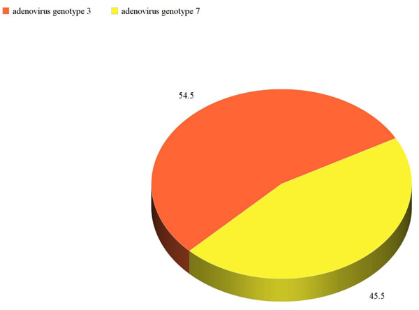
Adenovirus was detected by PCR for fiber gene in 11% with genotype 3 in 6 samples (54.5%) and genotype 7 in 5 samples (45.5%) positive for adenovirus by Multiplex PCR., (Fig. 1).
The main presenting symptoms and signs in children with confirmed adenovirus detected by PCR were cough, fever, wheezing, and croups (90.9%, 81.1%, 63.6%, and 63.6%, respectively). However, there was statistically insignificant difference in clinical symptoms as regards cough, fever, croup, dyspnea, dry rales, and moist rales (P=1.00, P= 0.5, P=0.8, P=1.00, P=1.00, P=0.5, respectively), while wheezy chest was significantly more frequent in children with confirmed adenovirus (63.6%) compared to children with no adenovirus (30.3%), P=0.04. The diagnosis in children with confirmed adenovirus was pneumonia in 72.7% and bronchitis in 27.7%. There were statistically insignificant differences in gender (P=1.00), and hematological parameters as regard to hemoglobin, lymphocytes count, neutrophil counts, and platelets counts between children with confirmed adenovirus and children negative to adenovirus by PCR (P=0.2, P=0.1, P=0.1, P=0.4 respectively) (Table 2).
| - | Children with Confirmed Adenovirus (n=11) |
Children Negative for Adenovirus (n=89) |
P |
|---|---|---|---|
| Age (mean± SD) months | 45.3 ± 8.7 | 45.8 ± 9.4 | - |
| Gender Male no (%) Female no (%) |
4 (36.4%) 7 (63.6%) |
31 (34.8%) 58 (65.2%) |
P=1.00 |
| Residence Rural no (%) Urban no (%) |
6 (54.5%) 5 (45.5%) |
64 (71.9%) 25 (28.1%) |
P=0.3 |
| Cough no (%) | 10 (90.9%) | 81 (91.1%) | P=1.00 |
| Fever no (%) | 9 (81.1%) | 61 (68.5%) | P=0.5 |
| Wheezing no (%) | 7 (63.6%) | 27 (30.3%) | P=0.04 |
| Croup no (%) | 7 (63.6%) | 47 (52.8%) | P=0.8 |
| Dyspnea no (%) | 3 (27.3%) | 24 (26.9%) | P=1.00 |
| Dry rales no (%) | 4 (36.4%) | 32 (35.9%) | P=1.00 |
| Moist rales no (%) | 2 (18.2%) | 28 (31.5%) | P=0.5 |
| Seizure no (%) | 1 (9.1%) | 1 (1.1%) | P=0.2 |
| Bronchitis no (%) Pneumonia no (%) Upper respiratory infections no (%) |
3 (27.3%) 8 (72.7%) 0 (0%) |
16 (17.9%) 69 (77.5%) 4 (4.5%) |
P=0.9 |
| Hemoglobin gm/dl (mean ± SD) | 14.0 ± 1.4 | 13.0 ± 2.2 | P=0.2 |
| Lymphocytes103/cmm (mean ± SD) | 4.4 ± 1.00 | 3.8 ± 1.1 | P=0.1 |
| Neutrophil103/cmm (mean ± SD) | 4.3 ± 1.00 | 5.2 ± 1.2 | P=0.1 |
| Platelets103/cmm (mean ± SD) | 163.7 ± 7.3 | 186.4 ± 81.2 | P=0.4 |
4. DISCUSSION
Adenoviruses are reported as common etiology of respiratory infections with a wide variance of occurrence from 2% up to 78% [19, 20]. In recent surveillance in Egypt about severe respiratory infections requiring hospitalization, children below 5 years represented 83% of patients [21].
In the present study, the mean age ± SD of children with adenovirus infection was 45.3 ± 8.7 months. This finding was similar to previous reports [22, 23]. This can be attributed to the immaturity of the immune systems of young children, which leaves them prone to more severe adenovirus disease.
The clinical characteristics of children with confirmed adenovirus were similar to children without adenovirus. It is known that clinical diagnosis has similar symptoms and signs with different viral etiologies in respiratory infections [24]. Therefore, there is a need to identify the specific viral pathogen. The use of conventional PCR for the detection of adenovirus with species and type-specific primers was proved to be an efficient and rapid method for the detection and typing of adenovirus associated with respiratory infections [25-27]. Accurate and rapid identification of HAdV infection in the respiratory tract not only avoids unnecessary antibiotic prescription but also prevents or inhibits HAdV-related outbreaks [25].
In the present study, this was performed by two steps molecular techniques. The first was by the determination of adenovirus by the use of PCR for fiber gene with common primers for adenovirus. The PCR was confirmed to be positive in 11 samples (11%). This result was consistent with previous reports with a range from 2% up to 13% [26, 27]. The differences in the determination of adenovirus in respiratory infections can be attributed to the difference of the studied population; the method used for the detection of adenovirus and even to the difference of the weather between countries as adenovirus is known to be more prevalent in hot dry weather [28].
The genotype determination of the positive samples for adenovirus revealed that adenovirus genotype 3 was determined in 6 samples (54.5%) and genotype 7 was present in 5 samples (45.5%) positive by Multiplex PCR. The common genotypes in respiratory infections in children were reported to be B (B3, B7, B21), C (C1, C2, C5, C6), and E (E4) [29, 30]. HAdV 2, 3, and 7 are the most prevalent species and are associated with severe pneumonia [31, 32]. In the present study, the diagnosis in children with adenovirus was pneumonia in 72.7% and bronchitis in 27.7%. This finding was online with previous studies [26, 27].
In general, the clinical characteristics of respiratory infections may vary with adenovirus and may vary upon the age of the patients and the immune status. Therefore, there is a requirement for an extensive study of adenovirus in respiratory infections in children with different ages and immune status.
CONCLUSION
The present study highlights the prevalence of adenovirus in Egyptian children with respiratory tract infections mainly in pneumonia with genotypes 3 and 7 as the predominant genotypes in those in children the finding which confirmed a worldwide trend of distribution of adenovirus infection in children.
ETHICS APPROVAL AND CONSENT TO PARTICIPATE
The study was approved by the Mansoura ethical committee- Institutional Research Board (IRB) with approval number R.19.09.637
HUMAN AND ANIMAL RIGHTS
No animals were used in the study. All reported humans were experimented in accordance with the ethical standards of the committee responsible for human experimentation (institutional and national), and with the Helsinki Declaration of 1975, as revised in 2008 (http://www.wma.net/en/20 activities/10ethics/10helsinki/).
CONSENT FOR PUBLICATION
Informed consent was obtained from the parents of each child.
AVAILABILITY OF DATA AND MATERIALS
The data supporting the findings of the article are available in the Figshare repository at https://figshare.com/articles/ revised_article3_doc/11829774
FUNDING
None.
CONFLICT OF INTEREST
The authors declare no conflict of interest, financial or otherwise.
ACKNOWLEDGEMENTS
Declared none.

