All published articles of this journal are available on ScienceDirect.

Effects of Culturing Factors and Reaction Conditions on the Synthesis of Gold Nanoparticles by Four Enterobacterial Species

Abstract
Introduction
Synthesis of gold nanoparticles (AuNPs) using enterobacteria confers many advantages, but the efficiency depends on the bacterial strains and culture conditions. Therefore, the selection of the most effective strains and the optimization of the culturing factors and reaction conditions are required.
Methods
Gold resistance and AuNP synthesis were evaluated across 105 enterobacterial strains. The AuNPs synthesized by the four selected strains were characterized using scanning electron microscopy (SEM). AuNP synthesis was improved by optimizing several parameters, including culture media, inducer concentrations, aeration, cultivation times, reaction temperatures, and incubation times.
Results
Fifty-six strains synthesized AuNPs at concentrations ranging from 7.56 ± 0.28 to 77.92 ± 3.92 μg/mL. The AuNPs synthesized by the four selected strains, including Citrobacter freundii ENTSF 1-3, Enterobacter cloacae ENTSF 8-1, Hafnia alvei ENTSF 15-1, and Morganella morganii SFTCBS1, appeared spherical to slightly polyhedral, with average sizes ranging from 22 to 28 nm. Lennox Luria-Bertani (LB) medium and static conditions were favourable culturing parameters for AuNP synthesis by all four strains. The most favourable reaction conditions varied among the four strains as follows: C. freundii ENTSF 1-3 (at 37°C for 72 h and 120 h); E. cloacae ENTSF 8-1 (at 55°C for 72 h and 120 h); H. alvei ENTSF 15-1 (at 37°C for 72 h and 120 h, as well as at 55°C for 24 h and 48 h); and M. morganii SFTCBS1 (at 55°C for 120 h).
Discussion
AuNP synthesis by the four selected strains offers several advantages, including a simple growth medium, a short cultivation time, a rapid reaction, convenient product harvesting, stable AuNPs, and no requirement for extra equipment.
Conclusion
This study determined the most suitable cultivation and reaction conditions to enhance the yield of AuNPs.
1. INTRODUCTION
Gold nanoparticles (AuNPs) are of great interest due to their unique and promising properties, including inertness, stability, biological activity, biocompatibility, high binding affinity, low toxicity, cell membrane penetration ability, apoptosis induction ability, interaction with light via surface plasmon resonance (SPR), optical properties, and electrical properties. AuNPs have been employed in a vast array of applications across multiple fields. AuNPs have been utilized in the detection of various biomolecules. In the medical field, AuNPs have been applied in diagnostics, anticancer therapy, photothermal therapy (PTT), photodynamic therapy (PDT), radiation therapy (RT), gene therapy, and immunotherapy. AuNPs have demonstrated therapeutic potential in antimicrobial, antidiabetic, antioxidant, and anti-inflammatory treatments. AuNPs have also been employed as carriers for drugs, antibiotics, nucleic acids, and proteins. In imaging processes, AuNPs have been used in X-ray photography and computed tomography (CT). In the agricultural field, AuNPs have been shown to promote plant growth and induce seed germination. In environmental applications, AuNPs have been used for air cleaning and water purification [1-3].
AuNPs display a variety of morphologies, encompassing differences in shape (e.g. sphere, oval, rod, cube, cage, shell, wire, branch, star, flower, bipyramid, triangle, pentagon, hexagon, and cluster) [2-4], size (approximately 2 to 200 nm) [2, 3], and colour (e.g. red, orange, brown, purple, and blue) [5]. The morphologies of AuNPs are crucial to their properties, including specificity, toxicity, cellular uptake ability, and cell membrane penetration ability [4]. The factors determining AuNP morphology include medium pH, reaction pH, temperature, time, metal salt concentration, trisodium citrate (Na3C6H5O7) concentration, and the ratio of trisodium citrate to chloroauric acid (HAuCl4) [2, 4, 6-8]. The reaction rates have been reported to be influenced by pH, temperature, salt concentration, and HAuCl4 concentration [2, 9].
Various approaches to generating AuNPs are categorized into three types: physical, chemical, and biological processes. Green technologies have increasingly gained popularity because of their advantages, including safety, eco-friendliness, high energy efficiency, product stability, and the avoidance of high temperatures and harsh chemicals. Numerous bacteria, fungi, yeasts, and algae have been successfully employed in the green synthesis of AuNPs.
Bacterial synthesis offers advantages because some bacteria are not affected by the presence of heavy metals, and their extracellular synthesis facilitates the purification process [2, 10]. The ability to synthesize AuNPs has been reported in various groups of bacteria as follows: (1) enterobacteria (e.g. C. freundii, Enterobacter xiangfangensis, Escherichia coli, Klebsiella pneumoniae, Proteus mirabilis, and Serratia marcescens) [11-16]; (2) marine bacteria (e.g.Bacillus marisflavi, Lysinibacillus odysseyi, Shewanella oneidensis, and Vibrio alginolyticus) [17-21]; (3) actinobacteria (e.g.Rhodococcus spp. and Streptomyces spp.) [22, 23]; (4) lactic acid bacteria (e.g.Lactobacillus acidophilus, Lactobacillus casei, and Streptococcus thermophilus) [9, 24]; and (5) other bacteria (e.g.Bacillus licheniformis, Bacillus subtilis, Pseudomonas aeruginosa, and Pseudomonas fluorescens) [9, 17, 25-28]. AuNP synthesis using enterobacteria confers the advantages of ease, rapidity, environmental friendliness, cost-efficiency, product uniformity, and feasibility for large-scale production [29, 30].
Due to the prominent ability of enterobacteria and their advantages in AuNP synthesis, this study was conducted to select the most effective strains, optimize the culturing factors and reaction conditions to maximize the yield of AuNPs synthesized by four enterobacterial species, and characterize the synthesized AuNPs using scanning electron microscopy (SEM).
2. MATERIALS AND METHODS
2.1. Enterobacterial Strains
A collection of 105 enterobacterial strains was established in our previous studies through the isolation of bacteria from 15 types of fresh seafood sourced in Thailand. All strains were identified to the genus level based on the sequence similarity of the 16S rRNA gene. Twenty-four strains were identified to the species level according to the VITEK 2 system. All strains were classified into nine genera: Citrobacter (20 strains), Enterobacter (55 strains), Hafnia (two strains), Klebsiella (six strains), Morganella (four strains), Providencia (12 strains), Salmonella (two strains), Serratia (two strains), and Yersinia (two strains) [31, 32].
2.2. Assessment of Gold Resistance Among 105 Enterobacterial Strains
The inocula, prepared by overnight cultivation of the strains in Lennox Luria-Bertani (LB) broth, were subcultured into Mueller-Hinton (MH) broth (Sigma-Aldrich, St. Louis, MO, USA) containing filter-sterilized HAuCl4·3H2O at concentrations of 19.5 mg/L (50 μM), 39 mg/L (100 μM), and 58.5 mg/L (150 μM) to obtain an initial concentration of 1.00 × 10⁵ colony-forming units (CFU)/mL. The bacterial cultures were grown at 37°C for 24 h. Turbidity and an optical density at 600 nm (OD600) above 0.1 indicated bacterial growth and, hence, gold resistance.
2.3. Determination of Concentrations of AuNPs Synthesized by 105 Enterobacterial Strains
The methodology for assessing AuNP synthesis was modified from the previously reported protocol [33]. The strains were cultivated in Lennox LB broth at 37°C for 36 h under static conditions. The cell-free supernatants were obtained by centrifugation of the cultures at 11,000 rpm for 10 min at 4°C in an Eppendorf 5804R centrifuge (Eppendorf, Selangor Darul Ehsan, Malaysia). The reaction mixtures were prepared by mixing 10 mL of supernatant with 10 mL of 1 mM HAuCl4 and then incubating the mixtures undisturbed at 37°C for 24 h and 120 h in the dark. The negative control was prepared using uninoculated medium instead of cell-free supernatant and served as a blank in the spectrophotometric method for determining AuNP concentration. AuNP synthesis was indicated by the colour changes of the reaction mixtures from light yellow to a diverse range of colours, including red, orange, brown, purple, and blue. The final colours were determined by the size, shape, and aggregation of the AuNPs [5, 10]. The ultraviolet-visible (UV-vis) spectra of the synthesized AuNPs were scanned using a NanoDrop 2000C spectrophotometer (Thermo Scientific, Waltham, MA, USA). The optical densities (ODs) at λmax of the reaction mixtures were quantified using a Cecil CE1011 spectrophotometer (Cecil Instruments, Cambridge, UK). A commercial AuNP suspension (Sigma-Aldrich, St. Louis, MO, USA) with the same λmax as the synthesized AuNPs was used for concentration quantification. A commercial AuNP suspension with an OD of 1.0 and a concentration of 35 µg/mL was serially diluted in Lennox LB medium to obtain concentrations ranging from 5 to 35 µg/mL. Lennox LB medium served as a blank in the spectrophotometric method. The OD was measured for each AuNP concentration. A standard curve was plotted using the OD values and AuNP concentrations. The concentrations of the synthesized AuNPs were quantified using the standard curve. All reactions were performed in triplicate.
2.4. Species Identification of the Selected Strains According to the VITEK 2 System
Enterobacterial strains from four genera were selected based on their highest concentrations of AuNPs synthesized. The selected strains were subjected to species identification using the Gram-Negative (GN) card of the VITEK 2 system version 07.01 (bioMérieux, Inc., Durham, NC, USA).
2.5. SEM Imaging of the Synthesized AuNPs
The reaction mixtures of the four selected strains were filtered through 0.45-μm pore size membranes (Sartorius Stedim Biotech, Gottingen, Germany). The filtrates were centrifuged at 11,000 rpm for 10 min at 4°C in an Eppendorf 5804R centrifuge (Eppendorf, Selangor Darul Ehsan, Malaysia) to obtain AuNP pellets. The AuNP pellets were washed three times and then dissolved in sterile ultrapure water (Invitrogen, Waltham, MA, USA). The morphologies of the synthesized AuNPs were characterized by Tescan Mira3 SEM (Tescan Orsay Holding, Brno-Kohoutovice, Czech Republic). The shapes and sizes of the synthesized AuNPs were analyzed with ImageJ software [34].
2.6. Determination of the Effects of Culturing Factors and Reaction Conditions on AuNP Synthesis by the Selected Strains
The inocula were prepared by cultivating the four selected strains in Lennox LB broth at 37 C for 36 h with shaking at 150 rpm. The inocula were adjusted to an initial concentration of 1.00 × 10⁵ CFU/mL for cultivation under conditions with varied culturing factors. The culturing factors were as follows: (1) culture media including Lennox LB (pH 7.0 ± 0.2) [35], brain heart infusion (BHI) (pH 7.4 ± 0.2) (Scharlab, Barcelona, Spain), and minimal medium M63 (pH 7.3 ± 0.2) [36]; (2) inducer concentrations (0, 20, and 50 μM HAuCl4); (3) static and aerated conditions (without shaking and with shaking at 150 rpm, respectively); and (4) cultivation times (6, 12, 24, 36, and 48 h). The varied reaction conditions were combinations of incubation temperatures (37°C and 55°C) and times (6, 24, 48, 72, and 120 h). Negative controls were performed using uninoculated media with the same concentrations of HAuCl4, rather than cell-free supernatants. These controls also served as blanks in the spectrophotometric method for determining AuNP concentration under each relevant condition. The AuNP concentrations were determined, as mentioned earlier.
2.7. Statistical Analysis
Data were analyzed using SPSS software version 19.0 (IBM Corp., Chicago, IL, USA). All experiments were performed in triplicate (n = 3). Results are expressed as the means ± standard deviations (SDs), supplemented by 95% confidence intervals (CIs). Significant differences among means were assessed using one-way or two-way analysis of variance (ANOVA), followed by Tukey’s HSD post hoc test for multiple comparisons. The magnitude of effects was quantified using partial eta squared (ηp2). A p-value < 0.05 was considered statistically significant. ANOVA summary outputs, including sums of squares, degrees of freedom (df), and F-statistics, were computed.
3. RESULTS
3.1. Gold Resistance Among 105 Enterobacterial Strains
Of the 105 enterobacterial strains screened for resistance to a gold compound, HAuCl4, 31 (29.5%), 7 (6.7%), and 11 (10.5%) strains were resistant at maximum concentrations of 50, 100, and 150 μM, respectively.
3.2. AuNP Synthesis by 105 Enterobacterial Strains
AuNP synthesis could be perceived from the color changes of the reaction mixtures from light-yellow to a diverse range of colors, including red, orange, brown, purple, and blue [5, 10]. These color changes were observed for 56 strains (53.3%). The color changes of the reaction mixtures due to the AuNP synthesis are shown in Fig. (1). The AuNP-producing bacteria were distributed in eight genera, including Enterobacter (29 strains), Citrobacter (nine strains), Providencia (seven strains), Klebsiella (three strains), Morganella (three strains), Serratia (two strains), Yersinia (two strains), and Hafnia (one strain). Fifty-six AuNP-producing strains synthesized extracellular AuNPs at concentrations ranging from 7.56 ± 0.28 to 77.92 ± 3.92 μg/mL. When comparing reaction incubation times of 24 h and 120 h, AuNP formation was observed in 34 strains at 120 h and in 22 strains at the earlier time point of 24 h. The four representative strains producing the highest AuNP concentrations within each genus were selected for further studies. The four selected strains and their concentrations of the synthesized AuNPs were Citrobacter sp. ENTSF 1-3 (56.64 ± 1.95 μg/mL), Enterobacter sp. ENTSF 8-1 (56.00 ± 3.84 μg/mL), Hafnia sp. ENTSF 15-1 (30.16 ± 1.68 μg/mL), and Morganella morganii SFTCBS1 (77.92 ± 3.92 μg/mL). All four strains synthesized AuNPs with a purple-red color and a λmax of 540 nm.
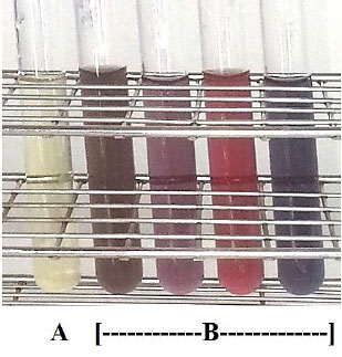
Color changes of reaction mixtures resulting from AuNP synthesis. The initial color of the reaction mixture was light yellow (A), and the final colors of the reaction mixtures containing AuNPs were brown, violet, red, and purple (B).
3.3. Species Identification of the Selected Strains According to the VITEK 2 System
Citrobacter sp. ENTSF 1-3, Enterobacter sp. ENTSF 8-1, and Hafnia sp. ENTSF 15-1 were previously identified at the genus level based on the alignment of partial 16S rRNA gene sequences [32]. These three strains were identified to the species level based on the VITEK 2 system as Citrobacter freundii ENTSF 1-3, Enterobacter cloacae subsp. cloacae ENTSF 8-1, and Hafnia alvei ENTSF 15-1, each with a probability of 99%. Morganella morganii subsp. sibonii SFTCBS1 was identified with a 99% probability using the VITEK 2 system in the previous study [31].
3.4. SEM Images of the Synthesized AuNPs
The SEM images (Fig. 2) depict the shapes and sizes of the AuNPs synthesized by four enterobacterial species. The synthesized AuNPs possessed spherical to slightly polyhedral shapes. The average sizes of the AuNPs synthesized by each strain were as follows: C. freundii ENTSF 1-3 (26.8 ± 14.4 nm); E. cloacae ENTSF 8-1 (27.8 ± 15.0 nm); H. alvei ENTSF 15-1 (22.1 ± 13.0 nm); and M. morganii SFTCBS1 (22.3 ± 13.2 nm).
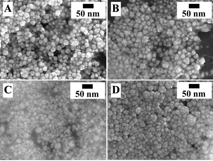
SEM images of AuNPs synthesized by enterobacterial strains. Spherical to slightly polyhedral AuNPs with average sizes ranging from 22 to 28 nm were synthesized by C. freundii ENTSF 1-3 (A), E. cloacae ENTSF 8-1 (B), H. alvei ENTSF 15-1 (C), and M. morganii SFTCBS1 (D).
3.5. Cultivation and Reaction Conditions Optimized for AuNP Synthesis by the Selected Strains
Factors promoting AuNP synthesis, including culture media, inducer concentrations, aeration, cultivation times, reaction temperatures, and incubation times, were optimized for the four selected strains. To evaluate the effect of culture media, the inocula of selected strains were grown in three culture media at 37°C for 36 h under static conditions. The AuNP concentrations were determined, as mentioned earlier. The concentrations of AuNPs synthesized by all four strains grown in three culture media are presented in Table 1. At both incubation times (24 h and 120 h), the AuNP concentrations were significantly highest for all four strains grown in Lennox LB.
| Bacterial Strain | Culture Medium | Reaction Incubation time (h) | AuNP Concentration (μg/mL) from a Culture Grown in Culture Medium (mean ± SD) | 95% Confidence Interval (CI) | Effect Size (ηp2) | p-value |
|---|---|---|---|---|---|---|
| C. freundii ENTSF 1-3 | Lennox LB | 24 | 38.12 ± 1.29h | [35.02, 41.23] | 0.99 | < 0.001 |
| 120 | 57.09 ± 2.42k | [51.07, 63.11] | 0.99 | 0.001 | ||
| E. cloacae ENTSF 8-1 | Lennox LB | 24 | 54.90 ± 2.86ijk | [47.78, 62.03] | 0.99 | 0.001 |
| 120 | 51.41 ± 0.69i | [49.67, 53.14] | 0.99 | < 0.001 | ||
| H. alvei ENTSF 15-1 | Lennox LB | 24 | 12.28 ± 0.88f | [10.08,14.47] | 0.99 | 0.002 |
| 120 | 29.69 ± 0.76g | [27.79, 31.59] | 0.99 | < 0.001 | ||
| M. morganii SFTCBS1 | Lennox LB | 24 | 76.85 ± 2.61l | [70.34, 83.36] | 0.99 | < 0.001 |
| 120 | 74.02 ± 2.35l | [68.18, 79.86] | 0.99 | < 0.001 | ||
| C. freundii ENTSF 1-3 | BHI | 24 | 0.33 ± 0.50a | [-0.93, 1.59] | 0.62 | 0.375 |
| 120 | 0.05 ± 0.09a | [-0.18, 0.30] | 0.57 | 0.423 | ||
| E. cloacae ENTSF 8-1 | BHI | 24 | 11.13 ± 0.18f | [10.66, 11.60] | 0.99 | < 0.001 |
| 120 | 8.76 ± 0.62ef | [7.21, 10.30] | 0.99 | 0.002 | ||
| H. alvei ENTSF 15-1 | BHI | 24 | 5.52 ± 1.03cde | [2.94, 8.09] | 0.98 | 0.012 |
| 120 | 0.05 ± 0.09a | [-1.76, 0.28] | 0.57 | 0.423 | ||
| M. morganii SFTCBS1 | BHI | 24 | 5.77 ± 0.86cde | [3.61, 7.93] | 0.99 | 0.007 |
| 120 | 2.43 ± 0.72abc | [0.63, 4.23] | 0.97 | 0.028 | ||
| C. freundii ENTSF 1-3 | M63 | 24 | 6.96 ± 1.16de | [4.06, 9.85] | 0.99 | 0.009 |
| 120 | 3.64 ± 1.10abcd | [0.88, 6.39] | 0.97 | 0.030 | ||
| E. cloacae ENTSF 8-1 | M63 | 24 | 0.53 ± 0.45a | [-0.59, 1.66] | 0.82 | 0.179 |
| 120 | 0.58 ± 0.53a | [-0.73, 1.90] | 0.80 | 0.197 | ||
| H. alvei ENTSF 15-1 | M63 | 24 | 1.43 ± 0.03a | [1.35, 1.50] | 0.99 | < 0.001 |
| 120 | 5.02 ± 0.49bcde | [4.90, 5.14] | 0.57 | < 0.001 | ||
| M. morganii SFTCBS1 | M63 | 24 | 1.57 ± 0.56ab | [0.18, 2.96] | 0.96 | 0.040 |
| 120 | 2.32 ± 0.16abc | [1.92, 2.71] | 0.99 | 0.002 |
Note: Values are expressed as means ± standard deviations (SDs) (n = 3). Superscript letters indicate significant differences (p < 0.05) determined by Tukey's HSD post hoc test, with shared letters indicating no significant difference. η p2 represents the partial eta squared (effect size).
To evaluate the effect of HAuCl4 as an inducer, the inocula of the selected strains were grown in Lennox LB broth without and with HAuCl4 at concentrations of 20 and 50 μM at 37°C for 36 h under static conditions. The AuNP concentrations were determined as mentioned earlier. The concentrations of AuNPs synthesized by all four strains grown in the absence and presence of HAuCl4 are presented in Table 2. The effect varied depending on the strain. The presence of 20 μM HAuCl4 yielded the significantly highest AuNP concentration from C. freundii ENTSF 1-3 at an incubation time of 120 h. The presence of 50 μM HAuCl4 yielded the significantly highest AuNP concentration from E. cloacae ENTSF 8-1 at an incubation time of 120 h. H. alvei ENTSF 15-1 did not resist 20 or 50 μM HAuCl4; hence, HAuCl4 could not promote AuNP synthesis by this strain. Although M. morganii SFTCBS1 resisted 20 and 50 μM HAuCl4, HAuCl4 was unable to promote AuNP synthesis by this strain.
| Bacterial Strain | HAuCl4 Concentration | Reaction Incubation Time (h) |
AuNP Concentration (μg/mL) from a Culture Grown in the Presence or Absence of HAuCl4 (mean ± SD) |
95% Confidence Interval (CI) | Effect Size (ηp2) | p-value |
|---|---|---|---|---|---|---|
| C. freundii ENTSF 1-3 | 0 μM | 24 | 40.25 ± 2.62cd | [33.72, 46.77] | 0.99 | 0.001 |
| 120 | 58.25 ± 3.62gh | [49.23, 67.26] | 0.99 | 0.001 | ||
| E. cloacae ENTSF 8-1 | 0 μM | 24 | 57.69 ± 0.87fgh | [55.52, 59.86] | 0.99 | < 0.001 |
| 120 | 51.12 ± 0.65e | [49.49, 52.74] | 0.99 | < 0.001 | ||
| H. alvei ENTSF 15-1 | 0 μM | 24 | 12.28 ± 0.87a | [10.11, 14.46] | 0.99 | 0.002 |
| 120 | 29.68 ± 0.76b | [27.79, 31.57] | 0.99 | < 0.001 | ||
| M. morganii SFTCBS1 | 0 μM | 24 | 78.56 ± 0.79ij | [76.57, 80.54] | 0.99 | < 0.001 |
| 120 | 72.64 ± 3.68i | [63.49, 81.78] | 0.99 | 0.001 | ||
| C. freundii ENTSF 1-3 | 20 μM | 24 | 54.97 ± 3.60fgh | [46.01, 63.93] | 0.99 | 0.001 |
| 120 | 101.06 ± 5.37k | [87.70, 114.42] | 0.99 | 0.001 | ||
| E. cloacae ENTSF 8-1 | 20 μM | 24 | 71.52 ± 1.05i | [68.90, 74.14] | 0.99 | < 0.001 |
| 120 | 72.40 ± 1.44i | [68.81, 75.99] | 0.99 | < 0.001 | ||
| H. alvei ENTSF 15-1 | 20 μM | 24 | ND | ND | ND | ND |
| 120 | ND | ND | ND | ND | ||
| M. morganii SFTCBS1 | 20 μM | 24 | 59.44 ± 0.56h | [58.04, 60.83] | 0.99 | < 0.001 |
| 120 | 53.36 ± 3.14fg | [45.53, 61.18] | 0.99 | 0.001 | ||
| C. freundii ENTSF 1-3 | 50 μM | 24 | 34.61 ± 2.27bc | [28.97, 40.25] | 0.99 | 0.001 |
| 120 | 50.08 ± 3.11ef | [42.33, 57.82] | 0.99 | 0.001 | ||
| E. cloacae ENTSF 8-1 | 50 μM | 24 | 54.08 ± 1.18fgh | [51.12, 57.03] | 0.99 | < 0.001 |
| 120 | 81.12 ± 2.08j | [75.95, 86.30] | 0.99 | < 0.001 | ||
| H. alvei ENTSF 15-1 | 50 μM | 24 | ND | ND | ND | ND |
| 120 | ND | ND | ND | ND | ||
| M. morganii SFTCBS1 | 50 μM | 24 | 44.56 ± 1.44de | [40.98, 48.14] | 0.99 | < 0.001 |
| 120 | 30.45 ± 2.73b | [23.65, 37.24] | 0.99 | 0.003 |
Note: Values are expressed as means ± standard deviations (SDs) (n = 3). Superscript letters indicate significant differences (p < 0.05) determined by Tukey's HSD post hoc test, with shared letters indicating no significant difference. η p2 represents the partial eta squared (effect size).
ND, not determined due to no bacterial growth.
To evaluate the effect of aeration, the inocula of selected strains were grown in Lennox LB at 37°C for 36 h under static and aerated conditions. The AuNP concentrations were determined, as mentioned earlier. The concentrations of AuNPs synthesized by all four strains grown under static and aerated conditions are presented in Table 3. Static conditions significantly promoted AuNP synthesis by all four strains at both reaction incubation times (24 h and 120 h).
| Bacterial Strain | Condition | Reaction Incubation Time (h) | AuNP Concentration (μg/mL) from a Culture Grown Under Static or Aerated Conditions (mean ± SD) | 95% Confidence interval (CI) | Effect Size (ηp2) | p-value |
|---|---|---|---|---|---|---|
| C. freundii ENTSF 1-3 | Static | 24 | 38.76 ± 0.91e | [36.49, 41.04] | 0.99 | < 0.001 |
| 120 | 56.79 ± 0.15g | [56.39, 57.18] | 0.99 | < 0.001 | ||
| E. cloacae ENTSF 8-1 | Static | 24 | 57.70 ± 0.86g | [55.56, 59.85] | 0.99 | < 0.001 |
| 120 | 43.73 ± 0.33f | [42.90, 44.56] | 0.99 | < 0.001 | ||
| H. alvei ENTSF 15-1 | Static | 24 | 10.74 ± 1.60b | [6.76, 14.72] | 0.99 | 0.007 |
| 120 | 29.85 ± 0.91d | [27.58, 32.11] | 0.99 | < 0.001 | ||
| M. morganii SFTCBS1 | Static | 24 | 78.56 ± 0.79hi | [76.57, 80.54] | 0.99 | < 0.001 |
| 120 | 75.33 ± 2.90h | [68.10, 82.56] | 0.99 | < 0.001 | ||
| C. freundii ENTSF 1-3 | Aerated | 24 | 1.36 ± 0.54a | [0.00, 2.71] | 0.95 | 0.050 |
| 120 | 18.81 ± 0.27c | [18.13, 19.48] | 0.99 | < 0.001 | ||
| E. cloacae ENTSF 8-1 | Aerated | 24 | 2.16 ± 0.31a | [1.37, 2.94] | 0.99 | 0.007 |
| 120 | 3.25 ± 0.34a | [2.39, 4.11] | 0.99 | 0.004 | ||
| H. alvei ENTSF 15-1 | Aerated | 24 | 1.44 ± 0.85a | [-0.68, 3.57] | 0.89 | 0.100 |
| 120 | 0.74 ± 0.32a | [-0.06, 1.55] | 0.94 | 0.058 | ||
| M. morganii SFTCBS1 | Aerated | 24 | 1.36 ± 0.45a | [0.24, 2.47] | 0.96 | 0.035 |
| 120 | 1.44 ± 0.18a | [0.98, 1.89] | 0.99 | 0.005 |
Note: Values are expressed as means ± standard deviations (SDs) (n = 3). Superscript letters indicate significant differences (p < 0.05) determined by Tukey's HSD post hoc test, with shared letters indicating no significant difference. η p2 represents the partial eta squared (effect size).
To evaluate the effect of cultivation time, the inocula of selected strains were grown in Lennox LB at 37°C for five cultivation times under static conditions. The AuNP concentrations were determined, as mentioned earlier. The concentrations of AuNPs synthesized by all four strains grown for five cultivation times are presented in Table 4. The significantly highest AuNP concentrations from the four selected strains at various cultivation times were as follows: C. freundii ENTSF 1-3 (12-h-old cultures at an incubation time of 120 h); E. cloacae ENTSF 8-1 (36-h-old cultures at an incubation time of 24 h); H. alvei ENTSF 15-1 (36-h-old cultures at an incubation time of 120 h); and M. morganii SFTCBS1 (36-h-old cultures at both incubation times).
| Bacterial Strain | Cultivation Time | Reaction Incubation Time (h) | AuNP Concentration (μg/mL) from a Culture Grown for a Cultivation Time (mean ± SD) | 95% Confidence Interval (CI) | Effect Size (ηp2) | p-value |
|---|---|---|---|---|---|---|
| C. freundii ENTSF 1-3 | 6 h | 24 | 4.50 ± 0.47abc | [3.32, 5.69] | 0.99 | 0.004 |
| 120 | 8.89 ± 0.47defg | [7.70, 10.07] | 0.99 | 0.001 | ||
| E. cloacae ENTSF 8-1 | 6 h | 24 | 8.33 ± 0.42cdefg | [7.27, 9.39] | 0.99 | 0.001 |
| 120 | 4.92 ± 0.64ab | [3.32, 6.51] | 0.99 | 0.006 | ||
| H. alvei ENTSF 15-1 | 6 h | 24 | 4.40 ± 0.32bc | [3.58, 5.21] | 0.99 | 0.002 |
| 120 | 1.32 ± 0.58a | [-0.12, 2.76] | 0.94 | 0.059 | ||
| M. morganii SFTCBS1 | 6 h | 24 | 12.20 ± 0.12g | [11.90, 12.51] | 0.99 | < 0.001 |
| 120 | 10.52 ± 0.36ef | [9.60, 11.43] | 0.99 | < 0.001 | ||
| C. freundii ENTSF 1-3 | 12 h | 24 | 44.72 ± 2.58n | [38.28, 51.15] | 0.99 | 0.001 |
| 120 | 68.80 ± 0.47s | [57.05, 80.54] | 0.99 | 0.002 | ||
| E. cloacae ENTSF 8-1 | 12 h | 24 | 10.68 ± 0.82fg | [8.64, 12.71] | 0.99 | 0.002 |
| 120 | 9.60 ± 1.25efg | [6.48, 12.71] | 0.99 | 0.006 | ||
| H. alvei ENTSF 15-1 | 12 h | 24 | 6.36 ± 1.39bcde | [2.90, 9.81] | 0.98 | 0.016 |
| 120 | 4.39 ± 0.84abc | [2.29, 6.49] | 0.98 | 0.012 | ||
| M. morganii SFTCBS1 | 12 h | 24 | 24.04 ± 0.24h | [23.63, 24.64] | 0.99 | < 0.001 |
| 120 | 33.07 ± 0.12jk | [32.77, 33.37] | 0.99 | < 0.001 | ||
| C. freundii ENTSF 1-3 | 24 h | 24 | 38.65 ± 1.73lm | [34.34, 42.96] | 0.99 | 0.001 |
| 120 | 61.83 ± 1.28qr | [58.65, 65.02] | 0.99 | < 0.001 | ||
| E. cloacae ENTSF 8-1 | 24 h | 24 | 35.12 ± 0.19kl | [34.64, 35.59] | 0.99 | < 0.001 |
| 120 | 32.64 ± 0.16j | [32.24, 33.03] | 0.99 | < 0.001 | ||
| H. alvei ENTSF 15-1 | 24 h | 24 | 9.03 ± 1.00defg | [6.60, 11.57] | 0.99 | 0.004 |
| 120 | 6.65 ± 0.76bc | [4.76, 8.54] | 0.99 | 0.004 | ||
| M. morganii SFTCBS1 | 24 h | 24 | 50.40 ± 0.55o | [49.02, 51.78] | 0.99 | < 0.001 |
| 120 | 62.16 ± 2.48r | [56.00, 68.33] | 0.99 | 0.001 | ||
| C. freundii ENTSF 1-3 | 36 h | 24 | 39.26 ± 1.45lm | [35.64, 42.89] | 0.99 | < 0.001 |
| 120 | 57.65 ± 1.37pq | [54.25, 61.05] | 0.99 | < 0.001 | ||
| E. cloacae ENTSF 8-1 | 36 h | 24 | 57.60 ± 0.88p | [55.41, 59.78] | 0.99 | < 0.001 |
| 120 | 50.80 ± 0.31o | [50.01, 51.58] | 0.99 | < 0.001 | ||
| H. alvei ENTSF 15-1 | 36 h | 24 | 11.09 ± 1.92g | [6.30, 15.88] | 0.98 | 0.010 |
| 120 | 31.77 ± 0.35jk | [30.88, 32.66] | 0.99 | < 0.001 | ||
| M. morganii SFTCBS1 | 36 h | 24 | 73.98 ± 1.44t | [70.39, 77.57] | 0.99 | < 0.001 |
| 120 | 75.97 ± 1.52t | [72.48, 79.76] | 0.99 | < 0.001 | ||
| C. freundii ENTSF 1-3 | 48 h | 24 | 5.07 ± 0.90abcd | [2.81, 7.32] | 0.98 | 0.010 |
| 120 | 10.93 ± 0.98g | [8.56, 13.30] | 0.99 | 0.003 | ||
| E. cloacae ENTSF 8-1 | 48 h | 24 | 30.29 ± 0.36ij | [29.39, 31.19] | 0.99 | < 0.001 |
| 120 | 41.30 ± 0.39mn | [40.32, 42.28] | 0.99 | < 0.001 | ||
| H. alvei ENTSF 15-1 | 48 h | 24 | 2.04 ± 0.68a | [0.35, 3.72] | 0.96 | 0.035 |
| 120 | 2.77 ± 0.79ab | [0.80, 4.74] | 0.97 | 0.026 | ||
| M. morganii SFTCBS1 | 48 h | 24 | 26.87 ± 0.24hi | [26.26, 27.48] | 0.99 | < 0.001 |
| 120 | 33.88 ± 0.24jk | [33.27, 34.48] | 0.99 | < 0.001 |
Note: Values are expressed as means ± standard deviations (SDs) (n = 3). Superscript letters indicate significant differences (p < 0.05) determined by Tukey's HSD post hoc test, with shared letters indicating no significant difference. η p2 represents the partial eta squared (effect size).
To evaluate the effect of reaction incubation temperatures and times, the inocula of selected strains were grown in Lennox LB at 37°C for 36 h under static conditions. The AuNP concentrations were determined, as mentioned earlier, except that the reactions were performed at 10 combinations of incubation temperature and time. The AuNP concentrations obtained from reaction incubation temperatures and times are presented in Table 5. The significantly highest AuNP concentrations from the four selected strains at various reaction temperatures and times were as follows: C. freundii ENTSF 1-3 (37°C for 72 h and 120 h); E. cloacae ENTSF 8-1 (55°C for 72 h and 120 h); H. alvei ENTSF 15-1 (37°C for 72 h and 120 h as well as at 55°C for 24 h and 48 h); and M. morganii SFTCBS1 (55°C for 120 h). ANOVA summary outputs, including sums of squares, degrees of freedom (df), and F-statistics, are presented in Table 6.
| Bacterial Strain | Incubation Time | Incubation Temperature |
AuNP Concentration (μg/mL) from a Reaction at an Incubation Temperature and Time (mean ± SD) |
95% Confidence Interval (CI) | Effect Size (ηp2) | p-value |
|---|---|---|---|---|---|---|
| C. freundii ENTSF 1-3 | 6 h | 37°C | 8.36 ± 0.31ab | [7.59, 9.14] | 0.99 | < 0.001 |
| 55°C | 18.08 ± 0.20cd | [17.58, 18.57] | 0.99 | < 0.001 | ||
| E. cloacae ENTSF 8-1 | 6 h | 37°C | 18.64 ± 0.15cd | [18.25, 19.02] | 0.99 | < 0.001 |
| 55°C | 25.96 ± 0.24efgh | [25.35, 26.56] | 0.99 | < 0.001 | ||
| H. alvei ENTSF 15-1 | 6 h | 37°C | 0.22 ± 0.35a | [-0.66, 1.11] | 0.61 | 0.388 |
| 55°C | 3.12 ± 0.48b | [1.91, 4.32] | 0.99 | 0.008 | ||
| M. morganii SFTCBS1 | 6 h | 37°C | 18.14 ± 0.16cd | [17.73, 18.56] | 0.99 | < 0.001 |
| 55°C | 27.33 ± 0.12efghi | [27.02, 27.63] | 0.99 | < 0.001 | ||
| C. freundii ENTSF 1-3 | 24 h | 37°C | 38.01 ± 0.26jk | [37.35, 38.67] | 0.99 | < 0.001 |
| 55°C | 21.08 ± 0.20def | [20.58, 21.57] | 0.99 | < 0.001 | ||
| E. cloacae ENTSF 8-1 | 24 h | 37°C | 54.26 ± 2.07m | [49.12, 59.41] | 0.99 | < 0.001 |
| 55°C | 65.76 ± 1.11n | [62.99, 68.52] | 0.99 | < 0.001 | ||
| H. alvei ENTSF 15-1 | 24 h | 37°C | 11.08 ± 0.59c | [9.60, 12.55] | 0.99 | 0.001 |
| 55°C | 34.04 ± 0.31hij | [33.26, 34.81] | 0.99 | < 0.001 | ||
| M. morganii SFTCBS1 | 24 h | 37°C | 75.92 ± 0.87o | [73.74, 78.09] | 0.99 | < 0.001 |
| 55°C | 101.28 ± 3.21p | [93.30, 109.25] | 0.99 | < 0.001 | ||
| C. freundii ENTSF 1-3 | 48 h | 37°C | 44.40 ± 1.83kl | [39.83, 48.96] | 0.99 | 0.001 |
| 55°C | 23.54 ± 0.52defg | [22.23, 24.85] | 0.99 | < 0.001 | ||
| E. cloacae ENTSF 8-1 | 48 h | 37°C | 57.14 ± 0.87m | [54.96, 59.32] | 0.99 | < 0.001 |
| 55°C | 76.74 ± 1.27o | [73.57, 79.91] | 0.99 | < 0.001 | ||
| H. alvei ENTSF 15-1 | 48 h | 37°C | 20.07 ± 0.68de | [18.36, 21.77] | 0.99 | < 0.001 |
| 55°C | 35.23 ± 0.15ij | [34.89, 35.66] | 0.99 | 0.001 | ||
| M. morganii SFTCBS1 | 48 h | 37°C | 74.89 ± 4.88o | [62.75, 87.03] | 0.99 | 0.001 |
| 55°C | 112.80 ± 5.44q | [99.28, 126.32] | 0.99 | < 0.001 | ||
| C. freundii ENTSF 1-3 | 72 h | 37°C | 55.43 ± 1.04m | [52.82, 58.04] | 0.99 | < 0.001 |
| 55°C | 24.42 ± 0.92defg | [22.11, 26.73] | 0.99 | < 0.001 | ||
| E. cloacae ENTSF 8-1 | 72 h | 37°C | 52.48± 2.17lm | [47.07, 57.88] | 0.99 | 0.001 |
| 55°C | 94.24 ± 4.15p | [83.92, 104.55] | 0.99 | 0.001 | ||
| H. alvei ENTSF 15-1 | 72 h | 37°C | 29.17 ± 0.44fghi | [28.07, 30.27] | 0.99 | < 0.001 |
| 55°C | 25.84 ± 0.53de | [24.50, 27.17] | 0.99 | < 0.001 | ||
| M. morganii SFTCBS1 | 72 h | 37°C | 74.40 ± 1.60o | [70.41, 78.38] | 0.99 | < 0.001 |
| 55°C | 115.36 ± 5.28q | [102.22, 128.49] | 0.99 | 0.001 | ||
| C. freundii ENTSF 1-3 | 120 h | 37°C | 58.00 ± 0.95mn | [55.62, 60.38] | 0.99 | < 0.001 |
| 55°C | 25.64 ± 0.59defgh | [24.16, 27.11] | 0.99 | < 0.001 | ||
| E. cloacae ENTSF 8-1 | 120 h | 37°C | 50.47 ± 1.12lm | [47.67, 53.28] | 0.99 | < 0.001 |
| 55°C | 98.20 ± 7.09p | [80.57, 115.82] | 0.99 | 0.002 | ||
| H. alvei ENTSF 15-1 | 120 h | 37°C | 30.32 ± 0.51ghij | [29.04, 31.61] | 0.99 | < 0.001 |
| 55°C | 7.62 ± 0.49ab | [6.39, 8.86] | 0.99 | 0.001 | ||
| M. morganii SFTCBS1 | 120 h | 37°C | 78.02 ± 6.64o | [61.53, 94.52] | 0.99 | 0.002 |
| 55°C | 131.19 ± 6.08r | [116.08, 146.30] | 0.99 | 0.001 |
Note: Values are expressed as means ± standard deviations (SDs) (n = 3). Superscript letters indicate significant differences (p < 0.05) determined by Tukey's HSD post hoc test, with shared letters indicating no significant difference. η p2 represents the partial eta squared (effect size).
| Experiment/Source | Sum of Squares | Degrees of Freedom (df) | Mean Square | F-value | Sig. (p-value) |
|---|---|---|---|---|---|
| AuNP concentrations obtained from the strains grown in three culture media | 44102.590 | 23 | 1917.504 | 1264.987 | < 0.001 |
| AuNP concentrations obtained from the strains grown in the absence and presence of HAuCl4 | 24401.450 | 19 | 1284.287 | 209.903 | < 0.001 |
| AuNP concentrations obtained from the strains grown under static and aerated conditions | 36203.372 | 15 | 2413.558 | 2430.057 | < 0.001 |
| AuNP concentrations obtained from the strains grown for five cultivation times | 61743.318 | 39 | 1583.162 | 977.074 | < 0.001 |
| AuNP concentrations obtained from the reactions performed at ten combinations of reaction incubation temperatures and times | 134733.494 | 39 | 3454.705 | 516.956 | < 0.001 |
4. DISCUSSION
All 49 strains capable of resisting at least 50 μM HAuCl4 belonged to six genera: Enterobacter (27 strains), Citrobacter (ten strains), Providencia (six strains), Klebsiella (two strains), Morganella (two strains), and Yersinia (two strains). A threshold concentration for gold resistance in bacteria has yet to be precisely defined. Instead, the threshold concentration varies across bacterial species, environments, and types of gold compounds tested. Even though AuNPs are viewed as promising agents for the treatment of multidrug-resistant bacteria due to their ability to both directly combat bacteria and deliver antibiotics, many bacteria possess mechanisms to overcome the toxicity of gold and can survive at high concentrations of gold compounds [37]. Gold-resistant bacteria and the concentrations at which they resisted have been reported in earlier studies, such as Acinetobacter sp. (20 μM AuCl3) [38], Arthrobacter sp. (10 μM AuCl3), Enterobacter sp. (10 μM AuCl3), Pseudomonas sp. (10 μM AuCl3), Serratia sp. (50 μM AuCl3), and Stenotrophomonas sp. (25 μM AuCl3) [39]. The four enterobacterial strains that synthesized the highest AuNP concentrations within their respective genera were selected. The four selected strains, namely C. freundii ENTSF 1-3, E. cloacae ENTSF 8-1, H. alvei ENTSF 15-1, and M. morganii SFTCBS1, synthesized AuNPs with spherical to slightly polyhedral shapes and average sizes ranging from 22 to 28 nm. Other Citrobacter and Enterobacter strains have been reported to synthesize AuNPs with similar morphologies. The C. freundii-derived AuNPs were spherical and polydisperse, with a size range of 30–60 nm [40]. The Enterobacter aerogenes-derived AuNPs were spherical and monodisperse, with a size range of 4–6 nm [30]. Of the 105 strains used in this study, 63 were previously classified as silver resistant based on their ability to grow in the presence of AgNO3 at a concentration exceeding 512 mg/L, and 22 synthesized silver nanoparticles (AgNPs) [41].
Statistical comparisons of AuNP concentrations were performed to determine the optimal culturing factors and reaction conditions for all four strains. Lennox LB medium and static conditions were the most suitable parameters, eliciting significantly higher AuNP synthesis by all four strains, whereas the most suitable inducer concentrations, culture ages, and reaction conditions were strain dependent. Among the four selected strains, M. morganii SFTCBS1 performed best in AuNP synthesis under the optimized parameters. The rationale that Lennox LB medium and static conditions promote AuNP synthesis is supported by previous studies. Bacterial AuNP synthesis is driven by various enzymes and metabolites. Bacterial metabolites acting as reducing agents in AuNP synthesis include aromatic compounds (e.g. prodigiosin), lipopeptides (e.g. surfactin), peptides, amines, amides, carboxylic acids, esters, alkanes, alkynes, phenols, and phosphates [42], and these metabolites are abundant in LB medium [43]. Nitrate-dependent reductase, NADPH-dependent reductase, and lactate dehydrogenase are key enzymes involved in AuNP synthesis [44, 45]. A medium containing glucose, peptone, yeast extract, and potassium nitrate (KNO3) at a specific ratio (1.5:1:0.35:0.35), whose composition is similar to that of Lennox LB medium, enhanced the nitrate-dependent reductase activity of B. licheniformis, thereby promoting the synthesis of AgNPs [46]. As nitrate reductase is negatively regulated by oxygen, anaerobic conditions and oxygen concentrations of 4% or lower were found to induce the synthesis of this enzyme, which was responsible for AgNP synthesis by E. coli [47-49]. The inducer HAuCl4 affects AuNP synthesis by liberating Au3+ ions for reduction to Au0 [17]. While HAuCl4 at concentrations of 0, 20, or 50 μM was most suitable for each strain in this study, 1 mM was the most suitable concentration for B. subtilis, E. coli, L. acidophilus, and S. thermophilus [9].
The effects of various parameters on AuNP synthesis have been determined in previous studies. The most promising parameters for AuNP synthesis by L. odysseyi included a ratio of HAuCl4 to bacterial extract of 1:9, a reaction temperature of 75°C, a reaction pH of 9.0, a bacterial extract concentration of 10%, and an HAuCl4 concentration of 1.0 mM [17]. Kikuchi et al. [24] attested that the ratio of bacterial cells to potassium gold (III) chloride (KAuCl4) concentration was crucial to AuNP synthesis by L. casei. AuNP synthesis was inhibited by an excess of either L. casei cells or KAuCl4. A combination of L. casei cells (1.0 g/L) and 0.25 mM KAuCl4 yielded AuNPs with λmax of 534 nm. Bacterial concentration influenced AuNP synthesis by P. aeruginosa, with 6 × 10⁸ cells/mL being the most effective concentration. Reaction conditions, including pH, temperature, and time, influenced the size and λmax of the P. aeruginosa-derived AuNPs. The reactions at 25°C for 48 h, 37°C for 24 h, and 42°C for 24 h yielded AuNPs with average sizes of 39.0, 26.0, and 36.7 nm, respectively [25].
The advantages of AuNP synthesis by the four selected strains were as follows: (1) A simple culture medium (Lennox LB) could be used for bacterial cultivation; (2) The maximum AuNP concentrations were achieved after short cultivation times (12–36 h); (3) AuNP synthesis was extracellular, thus offering easy and convenient product harvesting; (4) The synthesized AuNPs were stable and had narrow size distributions; (5) The reactions occurred within short periods; (6) Static conditions were preferred for bacterial cultivation, thus eliminating the requirement for aeration equipment and associated costs; (7) The reactions could occur at temperatures of 37°C and 55°C, thus eliminating the requirement for methods and apparatus designed for high or boiling temperatures; and (8) Condition optimization yielded the maximum AuNP concentrations of 101.06 ± 5.37, 98.20 ± 7.09, 35.23 ± 0.15, and 131.19 ± 6.08 μg/mL from C. freundii ENTSF 1-3, E. cloacae ENTSF 8-1, H. alvei ENTSF 15-1, and M. morganii SFTCBS1, respectively.
In this study, the synergistic effects of various factors, including medium type, inducer concentration, aeration, culture age, and reaction conditions, were demonstrated. AuNP synthesis by the four selected strains provides novel and promising alternatives to those achieved using other bacterial strains. The gold resistance characteristics of the selected strains would facilitate their survival under high concentrations of gold compounds and contribute to a higher yield of the synthesized AuNPs. Faster reactions were obtained with the selected strains from four genera than with Aeromonas, Arthrobacter, Comamonas, Elizabethkingia, Pseudomonas, and Streptomyces, which required 48 to 120 h [50, 51]. Compared to various bacterial species that synthesized AuNPs with different shapes and sizes, the four enterobacterial species synthesized AuNPs with spherical to slightly polyhedral shapes and average sizes ranging from 22 to 28 nm. This AuNP morphology has ideal properties for drug delivery and photothermal therapy. These AuNPs exhibit significantly higher uptake in radiation therapy for cancer treatment than smaller AuNPs and significantly higher tumour extravasation than larger AuNPs. This AuNP morphology provides longer persistence in the bloodstream, more precise malignant cell internalization, and the highest concentration and permanence within the target cell, as well as excellent thermal stability and high photothermal conversion efficiency [4, 52-54].
5. STUDY LIMITATIONS
The limitations of this study arise from the lack of the following information: (1) biological mechanisms supporting the effects of the examined parameters on AuNP synthesis; (2) determination of the effects of parameters on AuNP morphology; (3) morphological control of the synthesized AuNPs across diverse enterobacterial genera; (4) determination of the thermal stability of enzymes responsible for AuNP synthesis; (5) scale-up and replicative efficiency; and (6) demonstration of the applications of the synthesized AuNPs.
CONCLUSION
Gold resistance and AuNP-synthesizing ability were found to be common characteristics among 105 seafood-associated enterobacterial strains. The four selected strains from different genera, namely C. freundii ENTSF 1-3, E. cloacae ENTSF 8-1, H. alvei ENTSF 15-1, and M. morganii SFTCBS1, synthesized AuNPs with λmax peaks at 540 nm, spherical to slightly polyhedral shapes, and average sizes ranging from 22 to 28 nm. This study determined the most suitable cultivation and reaction conditions to enhance the yields of AuNPs synthesized by these strains. AuNP synthesis by these enterobacterial strains confers many benefits and therefore holds potential for future applications.
AUTHORS’ CONTRIBUTIONS
Both authors contributed equally to this article P.N. and N.P.: Conceived and designed the study, performed the experiments, analyzed the data, and wrote the manuscript.
LIST OF ABBREVIATIONS
| AuNPs | = Gold Nanoparticles |
| BHI | = Brain Heart Infusion |
| CFU | = Colony Forming Unit |
| Lennox LB | = Lennox Luria-Bertani |
| OD | = Optical Density |
| SEM | = Scanning Electron Microscopy |
AVAILABILITY OF DATA AND MATERIALS
All data generated or analyzed during this study are provided in this published article.
FUNDING
This work was financially supported by the Silpakorn University Research, Innovation and Creativity, Thailand, Administration Office under Grant SURDI 60/01/20.
ACKNOWLEDGEMENTS
Declared none.