All published articles of this journal are available on ScienceDirect.

Gene Editing for Tuberculosis using CRISPR-Cas Technology

Abstract
Advances in genome editing technologies are simplifying the tedious, laborious work in research. Recent advancements have introduced innovative genome editing techniques, such as the Clustered Regularly Interspaced Short Palindromic Repeat (CRISPR) and CRISPR-associated protein (Cas) system, which have proven to be powerful tools in genome editing. Unlike previous methods, CRISPR-Cas is highly specific and capable of gene knock-in, knock-out, or knockdown, which aids researchers. Such a tool is valuable for diagnosis, treatment, and vaccine production against life-threatening, mutating, and drug-resistant diseases such as Tuberculosis (TB).
Mycobacterium Tuberculosis (Mtb) and other members of the Mycobacterium Tuberculosis Complex (MTBC) are notorious and slow-growing bacteria; hence, they are difficult to tackle. Different CRISPR-Cas systems, for example, Cas9, Cas12a, Cas12b, and Cas13a, are the types being used in the diagnosis of Mycobacterium tuberculosis.
We have attempted to provide an overview of CRISPR-Cas technology and its application in tuberculosis. The literature was searched across platforms like PubMed, Google Scholar, Web of Science, SciELO, and Scopus. The keywords that were used to search the related literature were 'Mycobacterium tuberculosis OR Tuberculosis,' ‘TB AND Diagnosis,' ‘TB Vaccines AND Treatment,' ‘Mycobacterium tuberculosis AND CRISPR-Cas,' ‘CRISPR-Cas system,' ‘CRISPR-Cas in TB diagnosis,' 'Cas9,' 'Cas12a,' 'Cas13,' 'gene editing mycobacteria,' and ‘CRISPRi Mycobacterium tuberculosis.' The approach to searching for and selecting articles for the review provided a comprehensive and well-supported analysis of the subject matter.
The review emphasizes the novel technology CRISPR-Cas system for diagnosis, treatment, and the development of new TB vaccines. Along with the applications, the review comprises traditional methods of diagnosis of TB and mechanisms and challenges related to CRISPR-Cas. Using the CRISPRi technology, new targets were found as promising novel drug targets. Vaccine development with the help of this tool is also mentioned in this review.
CRISPR-Cas technology has emerged as a powerful tool for advancing TB research. Cas9, Cas12a, Cas12b, and Cas13 have demonstrated high specificity and sensitivity in detecting Mtb, even in samples with low bacterial loads. Moreover, CRISPR-mediated gene silencing has enhanced our understanding of the virulence mechanisms of the bacterium, facilitating the identification of novel therapeutic targets for designing new anti-TB drugs.
Tuberculosis continues to pose a major global health challenge due to the persistence of the pathogen and the emergence of drug-resistant strains. CRISPR-Cas technology offers innovative solutions, and its applications extend beyond diagnostics to include drug discovery, therapeutic development, and vaccine research. Thus, CRISPR-Cas systems can significantly accelerate efforts toward effective control and eradication of tuberculosis.
1. INTRODUCTION
Mycobacterium Tuberculosis (Mtb) is a member of the MTBC family and is among the most notorious bacteria causing TB. TB is an airborne infection that mainly affects the lungs and results in Pulmonary Tuberculosis (PTB). Apart from PTB, this bacterium is linked to several human illnesses, including metabolic syndromes, immunological disorders, and pulmonary problems, according to mounting data [1]. According to the World Health Organization (WHO) Report 2024, globally, 10.8 million cases of TB were documented, of which 8.2 million cases were newly diagnosed with TB [2]. Such increasing cases of TB pose a threat to global health. The issue is further aggravated by the rise in incidents of antimicrobial resistance to over-the-counter antibiotics [2]. It is therefore necessary to eliminate or manage this infection with the aid of developing technologies. Historically, TB was diagnosed based on patient symptoms such as hemoptysis (blood in the cough). In 1890, at his Berlin conference, Robert Koch proposed tuberculin as a medication for the disease, though it was later rejected as a treatment and instead suggested for diagnosis [3]. Later, in 1908, Charles Mantoux created an intradermal injection of tuberculin using a syringe and cannulated needle. In the 1930s, Florence Seibert of the Phipps Institute at the University of Pennsylvania produced a Pure Protein Derivative [PPD] in its present form after conducting several investigations [4]. As studies into the disease increased, it was diagnosed using various techniques, including staining [Ziehl-Neelsen Carbol Fuchsin], culture methods [Lowenstein-Jensen solid media], and X-ray examination of cavitary lesions. Increased knowledge of tuberculosis led to the introduction of immunological techniques for diagnosis, such as the enzyme-linked immunosorbent spot assay for cells that produce interferon-gamma [5]. At present, with the tests mentioned above, WHO also recommends tests like the molecular line probe assay and GeneXpert, which are valuable for detecting specific genetic markers associated with drug resistance, particularly in cases of MDR-TB or XDR-TB. With increasing technological advances, Whole Genome Sequencing [WGS] has also become a choice for TB diagnosis [6].
Once the disease has been diagnosed, medication is required to treat it. Before streptomycin was first developed as a therapy, earlier therapies for this illness, such as the royal touch and the sanatorium cure, were practiced [7]. The introduction of streptomycin altered the therapy paradigm, followed by rifampicin and isoniazid [8, 9]. With the discovery of new drugs and their combination, the fatality rate has decreased. According to the recommendation of the WHO, the initial treatment includes the first-line drugs, such as Isoniazid [INH], Rifampicin [RIF], Pyrazinamide [PZA], and Ethambutol [EMB], for 6 months. In the case of Multidrug-Resistant-TB [MDR-TB] or Rifampicin-Resistant-TB [RR-TB], the patient is treated with the second-line injectable drugs such as Fluoroquinolones [Ofloxacin [Ofl], Levofloxacin [LVX], and Moxifloxacin [MFX]] and Aminoglycosides [Amikacin [Am], Kanamycin [Kan], and Capreomycin [Cap]]. New therapy for MDR/RR-TB, known as BPaLM, consists of bedaquiline [B], pretomanid [Pa], linezolid [L], and moxifloxacin [M] for 6 months, and for those with pre-XDR-TB, the regimen can be taken without moxifloxacin [BPaL] for 9 months. [6]. Though the WHO recommends a variety of antibiotic regimens, drug-resistant TB treatment is a clinical conundrum. Therefore, it is imperative to find newer avenues and targets for intervention and mitigation.
Current scientific discoveries have opened new possibilities for diagnosing and treating tuberculosis. The CRISPR array, previously only employed as a phylogenetic marker in TB, now uses different Cas systems for diagnosis, new treatments, and enhancement of TB research [10]. The CRISPR-Cas system, which is a component of the immune system of prokaryotic cells, guards them against foreign genetic material. This system consists of two major components: the CRISPR locus, which is transcribed and subsequently cleaved to produce short CRISPR RNAs [crRNAs], and the Cas proteins, which are encoded alongside [11]. The CRISPR locus has spacer sections that contain the foreign genetic material that must be cleaved, as well as palindromic repeats after each spacer sequence, whereas Cas proteins are a kind of nuclease that cleaves the foreign nucleotide sequences at certain points [12]. This review focuses on the detection and novel drug development to eliminate TB. The review will also discuss the challenges concerning CRISPR-Cas technology in diagnosis and treatment.
2. METHODOLOGY
This literature review aims to compile existing knowledge, highlighting current research and advancements in using CRISPR-Cas9 technology for diagnosing and treating tuberculosis. The review involved searching for relevant articles on various academic databases like PubMed [https://pubmed.ncbi.nlm.nih.gov/], Google Scholar [https://scholar.google.com/], SciELO [https://www.scielo.org/en], Web of Science [https://mjl. clarivate.com/home], and Scopus [https://www.scopus.com /sources.uri?zone=TopNavBar&origin=searchbasic] using specific search terms like 'Mycobacterium tuberculosis OR Tuberculosis', ‘TB AND Diagnosis', ‘TB Vaccines AND Treatment', ' Mycobacterium tuberculosis AND CRISPR-Cas', ‘CRISPR-Cas system', and ‘CRISPR-Cas in TB diagnosis’, ‘Cas9’, ‘Cas12a’, ‘Cas13’, 'gene editing mycobacteria', and ‘CRISPRi Mycobacterium tuberculosis'. Additionally, reference lists from the identified articles and reviews were checked, and a forward and backward citation search was conducted to find additional relevant papers. No date restrictions were applied to the search, and all the relevant data were extracted. While extracting the peer reviews, articles, and reports, only the papers with the mentioned keywords were analyzed, whereas non-English papers, non-TB pathogens, and preprints were excluded in this review. High-impact papers were analyzed and thoroughly reviewed.
3. TRADITIONAL METHODS OF DIAGNOSIS OF TB DISEASE
Before diagnosis, it is necessary to understand how a person gets infected. Once the person gets infected with TB, they start showing signs and symptoms related to it, but in some cases, it is asymptomatic. As Mtb can infect any part of the body, the signs and symptoms depend on the site of the infection. In general, symptoms observed include fever, night sweats, and weight loss [13]. Understanding the symptoms and signs is important. The gold-standard diagnostic tool for detection is radiological examination [e.g., X-ray], which is preceded by microbiological investigations [14, 15]. Mantoux Tuberculin Skin Test [TST], Interferon γ Release Assay [IGRA], and, when indicated, Nucleic Acid Amplification [NAA] testing of sputum are done to confirm the disease [14–17].
3.1. Radiological Examination
The chest X-ray of a TB patient may show a caseous mass in the lungs. To further confirm the diagnosis, other tests are done to avoid misdiagnosis. Cases like Solitary Fibrous Tumours of the Pleura [SFTP] show the same ball-like structure as in TB, which is a common misinterpretation. Hence, radiological examination is not specific to tuberculosis [18].
3.2. Microbiological Investigations
The microbiological investigation includes culturing and staining processes where the TB bacteria are grown and observed. These bacteria grow slowly and require 6-8 weeks. Other bacteria in the sample grow much faster and contaminate the culture. Due to this, Ziehl-Neelsen staining becomes challenging. Even if the staining is done successfully, it does not indicate anti-TB drug resistance. Hence, it becomes difficult to detect TB when the number of bacteria in the sample is low, whereas culturing it becomes strenuous. Therefore, culturing TB bacteria is time-consuming and significantly prolongs the diagnosis [19].
3.3. Mantoux Tuberculin Skin Test [TST]
The TST is the most widely used, given its feasibility and low cost. The TST is performed on the forearm of the person suspected of TB. A PPD [tuberculin] is injected in the forearm, producing an elevated skin reaction, i.e. wheal. After injecting the tuberculin, the area is measured at 48-72 hours. If the person has TB, the area shows inflammation. The disadvantage related to this test is that it cannot differentiate between latent infection and active TB and fails to diagnose intraocular TB [20]. This test also yields false-positive results with BCG vaccination, which is a problem for newly vaccinated infants [19]. It fails to detect the disease in immunosuppressed and extrapulmonary patients [21].
3.4. Interferon Gamma Release Assay [IGRA]
The IGRA test is more specific than the TST but has its fair share of disadvantages. Similar to TST, this test also fails to distinguish between latent and current infection. It does not show any evidence of infection in immunosuppressed individuals [20].
3.5. Nucleic Acid Amplification Test [NAAT]
According to the WHO, nucleic acid amplification tests are highly recommended and have overcome some drawbacks of previous tests. This test includes Xpert MTB/RIF, Xpert MTB/RIF Ultra, Truenat, Genotype MTBDRplus, and Genotype MTBDRsl [2].
Xpert MTB/RIF and Xpert MTB/RIF Ultra: These tests are cartridge-based and include reagents and primers for Mtb resistance genes. The sample is liquefied before being placed into the cartridge. After loading, the entire cartridge is inserted into the machine [22]. These samples are detected for 81 core rpoB gene panels, including IS6110 and IS1081 for Mtb detection. Various versions, such as Xpert XDR, can identify types of Mtb drug resistance [17]. As with all other tests, this test also has certain limitations, such as the need for costly maintenance and electricity, and it shows no resistance in the case of poly-resistant TB [23]. Along with these limitations, it has suboptimal sensitivity and a high rate of false positive results, and it requires experience in handling samples [24]. Another issue is that cartridge shelf life is only 18 months, which makes it difficult for low-income nations to manage the cost [25].
Truenat: A chip-based, real-time PCR platform for TB diagnosis that is a portable and battery-operated machine. In this test, the sample is liquefied, and DNA extraction is performed. The extracted DNA is then mixed with the reagents of Truenat and loaded onto the chip, and real-time results are observed. Other versions of this test are Truenat MTB, Truenat MTB Plus, and Truenat MTB-Rif-Dx for the detection of Mtb as well as resistance genes [26]. The issue related to this test is that any mutation that occurs within the resistant gene is not detected through this test. Another disadvantage of this test is the viability of the bacilli, which can hinder the treatment process.
Genotype MTBDRplus and Genotype MTBDRsl: The test consists of three important steps, including DNA extraction, multiplex PCR amplification, and reverse hybridization. Reverse hybridization is performed on a special strip that contains zones where hybridization occurs. The associated disadvantages include a low bacillary count and the need for appropriate expertise for the test [27]. In a nutshell, all the above test shortcomings, viz., sensitivity, specificity, high maintenance, cost, expertise, bacillary count, and appropriate space, are the most important criteria to carry out the test and get appropriate results.
4. CRISPR-Cas TECHNOLOGY
The CRISPR-Cas system protects bacteria and archaea against invading conjugative plasmids, transposable elements, and viruses [28]. Jennifer Doudna and Emmanuelle Charpentier, with their discovery related to the CRISPR-Cas9 system as a gene editing tool, made genetic editing more precise and efficient [28]. As depicted in Table 1, there are two classes of the CRISPR-Cas system, i.e., Class 1 and Class 2, and each class is further divided into different types and subtypes [29, 30]. Mycobacterium tuberculosis contains a Class 1, type III-A CRISPR-Cas10 system as its defence system that targets foreign nucleic acids of plasmids as well as phages [31–33]. This type exhibits three enzymatic activities to cleave foreign nucleic acids [34–36].
| Characteristics | Type I | Type II | Type III | Type IV | Type V | Type VI |
|---|---|---|---|---|---|---|
| Class | Class 1 | Class 2 | Class 1 | Class 1 | Class 2 | Class 2 |
| Cas protein with nuclease activity | Cas3 and Cas7 | Cas9 | Cas10 | Csf2(Cas5), Csf3(Cas7) |
Cas12 | Cas13 |
| Target nucleic acid | DNA | DNA | DNA-RNA | Unknown | DNA | RNA |
| Cleaving pattern | ssDNA | dsDNA breaks blunt ends | Binds RNA to cleave |
Unknown | dsDNA breaks staggered ends | RNA |
| Subtypes | A-E, F1, F2, F3 and G | A to C | A to F | A to E | A - I, K, U and CRISPR- CasΦ |
A to B |
| References | [82–85] | [86–94] | [82–85] | [82,83,95–97] | [86,87,90,91,94,98,99] | [86,87,90,91,94,100,101] |
4.1. Mechanism of the CRISPR-Cas System
The CRISPR-Cas system in prokaryotes operates through three stages: adaptation, crRNA biogenesis/expression, and target interference, shown in Fig. (1) [37].
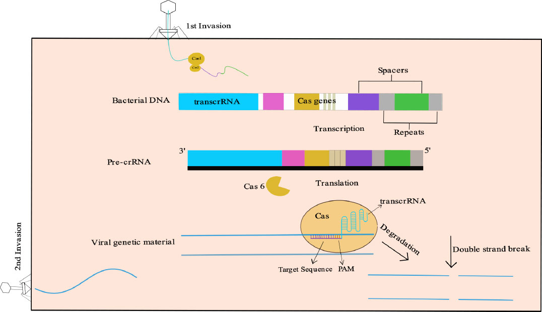
General mechanism of the CRISPR-Cas system [37].
- Adaptation: When a prokaryotic cell encounters foreign genetic material, such as a virus or plasmid, a small segment of it is cut and integrated into the CRISPR array in the spacer region called a protospacer. This process is facilitated by the Cas1 and Cas2 proteins, which are present in most CRISPR classes, except for type III C, D, and type IV [37, 38]. Although some research articles advocate a Reverse Transcriptase [RT]-fused-Cas1 protein in the type III-B system of Marinomonas mediterranea [39], in 2022, Aviram et al.’s study on the type III-A CRISPR-Cas system revealed that the adaptation system is RT-free [40]. More research is needed on the Class 1 CRISPR-Cas system to study its adaptation system. After the cleavage of foreign genetic material that integrates into the CRISPR array, the prokaryotic cell is allowed to “memorize” the invader's genetic information. It enables the bacterial immune system to recognize and mount a response against the same invader if it reappears. Some CRISPR types may use ancillary proteins to aid in the adaptation process [41].
- crRNA biogenesis/expression: The CRISPR array is transcribed into a precursor RNA transcript [pre-crRNA] and contains the spacers derived from the foreign genetic material. This precursor transcript is further processed to produce mature CRISPR RNAs [crRNAs] that carry the specific sequence information of the invader. The maturation of pre-crRNA into mature crRNA is mediated by specific proteins. For example, in class 1 CRISPR systems, the cas6 gene plays a major role in this process, while in type II systems, cas6 is absent and RNase III takes on the role. In type V systems, the cpf1 gene is involved in both adaptation and crRNA biogenesis/expression [41].
- Target interference: The mature crRNA serves as a guide to locate the specific genetic material of the invading element. Class 1 CRISPR systems utilize multiple effector proteins to mediate interference, while class 2 systems typically require only a single Cas protein to carry out this process. Types II, V, and VI CRISPR systems recognize a PAM sequence, a short specific sequence near the target site. The PAM sequence helps prevent the CRISPR system from mistakenly targeting the host's DNA [41].
Once the target is located, the interference stage involves the degradation or cleavage of the foreign genetic material. Cas3 protein, for example, plays a role in the cleavage of the targeted sequence. Additionally, the Cas10 protein has a dual function in target cleavage, binding both the crRNA and the target. Other proteins, such as the Csm and Cmr complexes, aid in the interference process for both DNA and RNA targets [37, 38, 41].
5. CRISPR-Cas-BASED DIAGNOSIS OF TUBERCUL OSIS
After the infection has occurred, it is very important to diagnose it before the disease worsens. In TB, due to slow growth, a lower bacterial load [paucibacillary TB], difficulty in sampling of Extrapulmonary TB [EPTB], and poor sensitivity and specificity of diagnostic tests lead to a delay in the diagnosis, which ultimately delays the treatment [42].
5.1. CRISPR-Cas9-based Detection
The most widely used CRISPR-Cas system is Cas9, which belongs to the class 2, type II nuclease group [43]. Tram et al. used Cas9 with multiplexed sgRNA that targets 52 genes associated with first-line and second-line drug-resistant genes along with IS6110 and IS1081 for identification of the MTBC strain. The diagnostic method utilized the CRISPR-Cas9 system for targeted enrichment followed by Illumina sequencing. After the target enrichment of the samples, the enriched samples were sequenced, which showed high-efficiency reads with a 104X depth, similar to normal culture whole-genome sequencing, which was enhanced to 2076X when the FLASH-TB technique was used. This technique, named Finding Low Abundance Sequences by Hybridization [FLASH], showed high sensitivity and specificity of 100% to Isoniazid [INH], Rifampicin [RIF], Ethambutol [EMB], Kanamycin [KAN], Amikacin [AMK], Moxifloxacin [MOX], Pyrazinamide [PZA], and Streptomycin [STR—only 90% specificity]. The expected Turnaround Time [TAT] is within 48 hours with a cost of $275. Although the method demonstrated high specificity and sensitivity, some small regions like ddn, fbiB, and fbiC had less than 10X coverage during sequencing [44]. Another CRISPR-Cas9-based detection technique is based on an electrochemical biosensor composed of a gold-modified screen-printed carbon electrode [SPCE/Au]. The electrode surface was immobilized with a ferrocene-labeled DNA probe [DNA probe-Fc] with a biotin group at the 3' end, with the help of streptavidin-biotin interaction, followed by blocking non-specific sites with Bovine Serum Albumin [BSA]. This detection tool focuses on mutations in the katG gene, which are responsible for INH resistance. The sensor with the immobilized ssDNA hybridizes with the target DNA to form dsDNA. This dsDNA is then recognized and cleaved by the CRISPR-Cas9 system with a gRNA designed to target codon 315, a common mutation site, with the mutation located near the 3' end of the gRNA target sequence for enhanced sensitivity. Due to the cleavage of the dsDNA, a ferrocene signal is produced as detected by Square Wave Voltammetry [SWV]. The biosensor was successfully able to detect DNA in the range of 101 to 106 Attomolar [aM]. This tool shows promising results for the diagnosis of low-load and resistant bacteria with a TAT of 6 to 7 hours [45].
Although both methods are highly specific and sensitive, they have high TAT, which is a drawback of these methods.
5.2. CRISPR-Cas12a-based Detection
CRISPR-Cas12a, also known as Cpf1, has proven to be a powerful tool for genetic editing. As a versatile genome editing tool with advantages like the self-processing of pre-crRNA into mature crRNA [46], the ability to target T-rich PAM sequences, and the generation of staggered DNA cuts with 5’ overhangs, shows an immense amount of application, especially in the field of detection [47, 48].
Zhang X and team made a detection kit for Mtb using CRISPR-Cas12a and Recombinase-Aided Amplification [RAA], which shows prominent specificity and sensitivity towards the identification of Mtb. Isothermal amplification was used along with three enzymes, namely recombinase [UvsX], Single-chain Binding Protein [SSB], and DNA polymerase [Klenow, having 3’-5’ exonuclease activity] [49]. The process was validated by analogizing with the gold standard technique of BACTEC 960 culture results.
The resultant Limit of Detection [LoD] was found to be 3.13 CFU/mL, or about 30 copies/µL. Compared with TAT, Xpert, loop-mediated isothermal amplification, and simultaneous amplification testing, TB-CRISPR took only 90 minutes. This method has overcome the TAT drawback; additionally, TB-CRISPR shows a specificity of 0.946 and a sensitivity of 0.883. The overall cost of the whole assay is about $6.90; compared to other molecular methods, this assay is less expensive [50].
Xiao et al. introduced another assay based on CRISPR-Cas12a using a combination of Graphene oxide-assisted multiplex recombinase polymerase amplification assay [GO-assisted multiplex RPA assay] that targets IS6110 and IS1081 in Mtb. The major parts of this assay are GO-assisted singleplex and multiplex RPA, CRISPR-Cas12a-based trans-cleavage, and Lateral Flow Biosensor [LFB]. The DNA from H37Ra was extracted, and GO-assisted singleplex and multiplex RPA were performed. In GO-assisted singleplex and multiplex RPA reactions, the extracted DNA was amplified using RPA. RPA is a well-known method for amplification with a characteristic of high sensitivity, but has a limitation of non-specific amplicon and primer dimer production. Therefore, in this method, GO was used to overcome this drawback, as mentioned by Munawar MA in 2022. After amplification, a trans-cleavage assay was performed with a complex IS6110-gRNA, IS1081-gRNA, and LbCas12a, making it a Cas12a-gRNA complex along with the fluorescence reporter molecule and RPA product. The fluorescence of the complex was detected. To evaluate this, a Lateral Flow Biosensor [LFB] was constructed using a Nitrocellulose [NC] membrane as a reaction platform. On the NC membrane, 3 different areas [conjugated pads] were coated with capture reagents. The first area was coated with Streptavidin-Gold Nanoparticles [SA-GNPs], which were close to the sample loading area; the second area, or the Control Line [CL], was coated with rabbit Anti-Fluorescein Amid [anti-FAM] antibody; and the third area, or the Test Line [TL], was coated with Biotin-Bovine Serum Albumin [biotin-BSA]. The treated samples were loaded along with the running buffer onto the LFB and allowed to flow through the coated NC, similar to a pregnancy kit, where the positive test shows two red lines on CL and TL. This kit was able to reach the LoD of 0.4 copies/µL. The overall process is completed in 60 minutes. It showed a sensitivity of 74.8% and a specificity of 100%. The kit has been approved by the Ethical Committee of Beijing Children’s Hospital, Capital Medical University [51]. As the mortality rate among people living with HIV [PLHIV] is high, it is essential to treat such people at the earliest. For adequate treatment of such patients, hypersensitive diagnosis is the most critical part for patients, including children. Hence, Huang et al. used the LFB assay for the detection of Cell-Free DNA [cfDNA] among these cohorts. This study strengthens and supports the detection kit for pulmonary and extrapulmonary TB detection [52].
With the advancement and a better understanding of the Cas12a system, Jia et al. introduced a novel detection technique. The latest amplification technique, Multiple Cross Displacement Amplification [MCDA] [53], along with CRISPR-Cas12a, was used to detect Mtb DNA. This technique consisted of three parts: 1. MCDA primer designing, 2. Standard MCDA assay, and 3. CRISPR-Cas12a-based detection. In MCDA primer designing, ten primers consisted of 2 replacement primers [F1, F2], 2 cross primers [CP1, CP2], and 6 amplification primers [C1, C2, D1, D2, R1, R2] that target the sdaA gene in Mtb. Between CP1 and gRNA [complementary to the P1 primer], a TT base was added as the PAM, as CRISPR-Cas12a is T-rich. A fluorescent label with FAM and Black Hole Quencher [BHQ1] was designed along with the primers. With the primers, an MCDA assay was performed with the samples and observed using real-time turbidity. Further, the detection was done using a CRISPR-Cas12a-based biosensing system on real-time PCR. The MTB-MCDA-CRISPR assay has 100% specificity for MTB detection. It can detect as low as 40 fg of MTB DNA. The assay has high sensitivity in clinical settings [54].
Since the TAT of the detection increases due to amplification, Wang and colleagues innovated a new technique by eliminating the PCR or isothermal amplification of the DNA. The detection technique consisted of immobilized CRISPR/Cas12a for DNA recognition and cleavage on Graphene Field-Effect Transistors [GFET] for electrical signal amplification. Graphene was used as the detection material, with the extracted DNA introduced onto the Cas12a-GFET. After binding of the Mtb DNA to the immobilized Cas12a-GFET, a signal is detected in 5 minutes with a sensitivity of 97.6% and a specificity of 98.6%. The limit of detection that can be detected by this method is about 2.42 x 10⁻¹⁸ M [55].
5.3. CRISPR-Cas12b-based Detection
Another subtype in class 2 type V is Cas12b, also denoted as C2c1. Cas12b has three parts: a nuclease with a single RuvC, a crRNA, and a transcrRNA. Advanced research in this type has revealed that Cas12b has significant applications in human genome editing [43, 56]. With effective targeting, Cas12b is also being used for Mtb detection.
With the surge of high-specificity, high-sensitivity, and cost-effective techniques, TB-Quick was introduced by Sam et al. Enhancing the previous findings and literature of HOLMES [a one-Hour Low-cost Multipurpose highly Efficient System] [57] and HOLMES.v2 [58], based on CRISPR-Cas12b, Sam et al. introduce a detection assay which is a combination of Loop-mediated Isothermal Amplification [LAMP] and CRISPR-Cas12b from Alicyclobacillus acidoterrestris [AacCas12b]. Amplification was performed using the most common gene, IS6110, as primers were made for targeting Mtb and using the LAMP kit, which contained three pairs of primers. Different temperatures were optimized to amplify the DNA extracted from types of samples, i.e., 65°C for 60 minutes for pulmonary samples like sputum/BALF DNA and for non-pulmonary samples like cfDNA, 65°C for 80 minutes, and 85°C for 10 minutes for the inactivation of the LAMP reaction [59]. The difference in amplification time is due to a higher concentration of bacilli as compared to non-pulmonary samples. A complete detection assay was performed after amplifying the 16 LAMP products, sgRNA, and Cas12b along with a fluorescence quenching probe [HEX-N12-BHQ1]. The detection was performed in a real-time PCR system, where the fluorescent signals were detected at 48°C for 10 minutes. The assay successfully detects samples containing DNA as low as 1.3 copies/µL. The overall time required for this detection is approximately 1-2 hours depending upon the sample. The specificity and sensitivity of the assay are approximately 95.2% and 86.8%, respectively [59]. In the search for a quick and feasible diagnosis of TB, Peng L and colleagues introduced an assay for fast and early TB diagnosis. TB One-Pot detection assay is a Cross-Priming-based Amplification [CPA] carried out using strand displacement polymerase [60, 61], along with Cas12b derived from Alicyclobacillus acidophilus [AapCas12b]. In this assay, as in the previously mentioned assays, Peng L has also used the highly conserved sequence of Mtb,i.e., IS6110 and IS1081, which helps target Mtb. The fluorescence was observed on a real-time system, and within 32 minutes, the results were obtained. The assay was also checked for cross-reactivity with 15 different Non-Tuberculous Mycobacteria [NTM] strains, namely M. simiae, M. parascrofulaceum, M. avium, M. intracellulare, M. paraintracellulare, M. kansasii, M. chelonae, M. abscessus, M. triplex, M. lentiflavum, M. paragordonae, M. gordonae, M. scrofulaceum, M. fortuitum, and M. colombiense, and six common respiratory pathogen strains: Aspergillus fumigatus, Cryptococcus neoformans, Pseudomonas aeruginosa, Escherichia coli, Klebsiella pneumoniae, and Staphylococcus aureus. Observation showed low fluorescence for these strains; on the other hand, the assay showed high fluorescence values for H37Rv and BCG. The assay specificity is 96.7%, and the sensitivity is 67.2%, whereas the LoD for TB One-Pot was 0.8 copies/µl. The combination of CRISPR-Cas12b and CPA has a limitation related to specificity and sensitivity. On comparison of cost with other available tests for Mtb detection, this is cost-effective and requires only $1.4 per test, which becomes affordable for the underprivileged [62].
5.4. CRISPR-Cas13-based Detection
CRISPR-Cas13, also known as the C2c2 system, is a subtype belonging to class 2 type VI, as explained previously. This RNA-guided RNA-targeting protein has tremendous applications in gene suppression in mammalian cells. Cas13 extracted from Leptotrichia wadei [LwaCas13a] provides the most efficient Cas for gene targeting in mammalian cell lines [63].
Ren et al. developed a diagnostic method using PCR and CRISPR-Cas13a that targeted IS1081, which is a conserved region in MTBC members. Targeting IS1081 showed high sensitivity by detecting the bacterial load of 1 target sequence copy/µl. The sensitivity of 97.4% and high specificity were compared with the gold standard [64].
One more technique for the detection of Mtb was done using CRISPR-Cas13a and RAA. IS6110 was used as a target with 100% specificity and no cross-reactivity with NTM. With a limit of detection of 10 copies/µl, the technique showed a sensitivity of 69%. The TAT was 1.5 hours with no false positive test [65].
By using the Cas13 collateral cleavage property, Sri Gowtham Thakku and colleagues created a new platform by improvising the SHERLOCK [Specific High Sensitivity Enzymatic Reporter UnLOCKing] technique [66] to WATSON [Whole Genome Assay using Tiled Surveillance of Nucleic acids] to detect cfDNA. The WATSON technique differs from the SHERLOCK technique in the amplification step, such that in the WATSON technique, the amplification is multiplex tiled-based and targets multiple targets, whereas in the SHERLOCK technique, the amplification is singleplex and targets only one target in the genome. This technique is highly applicable for samples with less concentration of DNA. For this technique, several primers were sequenced for compatibility using an iterative, heuristic search algorithm. After the primers were designed, all 18 crRNAs were synthesized by in vitro transcription. DNA from Mtb, NTM, and blood samples containing cfDNA was extracted, and a multiplex PCR amplification was performed. The reaction was performed with fluorescent-labelled Cas13, 18 pooled crRNA, and RPA reaction samples. The assay showed a sensitivity of 91% and a specificity of 89%. A Drop Array and a lateral flow assay were performed to confirm the test [67].
With an increasing need for early detection, specific and sensitive diagnostic assays are significant. Diagnosis of TB becomes challenging due to sample contamination, limited sample quantity, reduced growth of the bacteria, interference of NTMs, high cost, etc. Hence, CRISPR-Cas-based diagnosis strengthens the process in all aspects. A tabular summary of all CRISPR-Cas detection is depicted in Table 2.
| Cas Type | Targets | Amplification |
Detection Readout |
Sensitivity | Specificity | TAT | Cost | LoD | Advantages | Limitations |
|---|---|---|---|---|---|---|---|---|---|---|
| Cas9 | 52 drug resistance genes, IS6110, IS1081 | CRISPR enrichment | Illumina sequencing | 100% (most drugs) | 100% (90% STR) | 48 hrs | $275 | 104X coverage | Drug resistance profiling | Long TAT, low coverage in some regions |
| Cas9 | katG codon 315 (INH resistance) | None | Square wave voltammetry | Detects 101-106 aM | N/A | 6-7 hrs | N/A | 101 aM | Ultra-sensitive for mutations | Single mutation only |
| Cas12a | IS6110 | RAA | Fluorescence | 88.3% | 94.6% | 90 min | $6.90 | 3.13 CFU/mL | Cost-effective, fast | Moderate sensitivity |
| Cas12a | IS6110, IS1081 | GO-assisted RPA | Lateral flow biosensor | 74.8% | 100% | 60 min | N/A | 0.4 copies/μL | Visual readout, simple | Lower sensitivity |
| Cas12a | sdaA | MCDA | Real-time PCR fluorescence | High clinical | 100% | N/A | N/A | 40 fg DNA | Ultra-sensitive | Complex primers |
| Cas12a | IS1081 | None | Graphene FET electrical | 97.6% | 98.6% | 5 min | N/A | 2.42×10⁻¹⁸ M | No amplification needed | Requires specialized equipment |
| Cas12a | cfDNA (PLHIV/children) | RPA | Lateral flow | N/A | N/A | N/A | N/A | N/A | EPTB/cfDNA suitable | Limited performance data |
| Cas12b | IS6110 | LAMP | Real-time fluorescence | 86.8% | 95.2% | 1-2 hrs | N/A | 1.3 copies/μL | Simple workflow | Temperature optimization needed |
| Cas12b | IS6110, IS1081 | CPA | Real-time fluorescence | 67.2% | 96.7% | 32 min | $1.40 | 0.8 copies/μL | Cheapest, fastest | Lower sensitivity |
| Cas13 | IS1081 | PCR | Collateral cleavage | 97.4% | High | N/A | N/A | 1 copy/μL | High sensitivity | Requires PCR |
| Cas13 | IS6110 | RAA | Collateral cleavage | 69% | 100% | 1.5 hrs | N/A | 10 copies/μL | No cross-reactivity | Lower sensitivity |
| Cas13 | Multiplex tiled cfDNA | Multiplex PCR | Fluorescence/drop array/LFA | 91% | 89% | N/A | N/A | N/A | Genome-wide cfDNA | Complex multiplexing |
6. CRISPR-Cas-BASED DRUG TARGETS FOR ANTI-TUBERCULOSIS DRUGS
Multidrug resistance is a major concern related to TB. The targets for new drugs were identified using two approaches: i) 'Target-to-drug' and ii) 'Drug-to-target.' To target genes, protein mimics essential for bacterial growth and survival were created, and different drug studies were carried out [68]. Another way to target genes involves using homologous gene recombination, phage transduction, and high-density transposon mutant libraries, or knocking down genes by targeting essential genes required for the transcription process. These identification methods fall under the 'Target-to-drug' approach [69].
In the ‘Drug-to-target’ approach, assay plates such as the microplate Alamar assay or fluorescent-based screening were used for checking cell proliferation against the drug, a process that was time-consuming [70]. CRISPR is not only valuable for discovering new drug targets but also for studying the synergistic or antagonistic relationships between existing tuberculosis drugs and genes. Gene editing in Mycobacterium with old techniques was laborious and costly. With this new CRISPR-Cas9 technique, it has become feasible to edit a single gene or multiple genes in an organism. Gene silencing in Mycobacterium has become easier with the help of sgRNAs, which have catalytically dead or deactivated nuclease activity. This particular CRISPR interference [CRISPRi] technique targets the gene of interest using the PAM sequence and blocks the RNA polymerase from transcribing the DNA [71].
Choudhary et al. utilized this technique to target the yidC, gyrA, and engA genes in Mycobacterium smegmatis. In Mtb, they targeted engA, ftsZ, clpP2, groEL1, groES, clpC1, and yidC genes to assess the efficacy of CRISPRi in both fast- and slow-growing MTBC members [72]. In a study, CRISPRi was used to screen whiB7, a transcriptional regulator, as a potential new drug target for the drug clarithromycin, which was repurposed to target the gene [73].
7. VACCINE PRODUCTION
CRISPR-Cas9 has become a time-efficient tool for vaccine development. Time-consuming and labor-intensive knockouts required for live attenuated vaccines have now been simplified with this technique, making it a one-step process. Moraes et al. created an auxotrophic recombinant BCG strain that expressed a LTAK63 adjuvant [a detoxified subunit of E. coli heat-labile toxin] without using antibiotic resistance markers. Using the CRISPR-Cas9 technique lysA gene in Mycobacterium bovis BCG was knocked out, creating a lysine auxotrophic strain [BCGΔlysA] that cannot grow unless supplemented with the amino acid lysine. The auxotrophic strain was then transformed with a mycobacterial vector carrying both the functional lysA gene [to restore growth] and the ltak63 gene. The ltak63 gene played the role of a selection marker, replacing the use of antibiotics. rBCGΔ-LTAK63 emerged as a promising TB vaccine candidate, showing reduced bacterial load in the mouse model [74].
8. CHALLENGES In CRISPR-Cas SYSTEM-BASED DIAGNOSIS AND TREATMENT OF TUBERCULOSIS
Advancements in molecular genetics have enhanced the field of diagnosis and treatment. Despite being an advanced technology for diagnosing and treating tuberculosis, CRISPR-Cas cannot fill a few gaps. The limitations related to the diagnosis lie in sample processing, with the long and extensive procedure and the instruments required to process the sample, which require trained personnel. CRISPR-Cas-based assays need to be performed at a very low temperature to avoid denaturation of the protein [75]. In underdeveloped and developing countries, such a diagnosis becomes problematic if not handled with caution and can lead to misdiagnosis. Another disadvantage of CRISPR-Cas-based treatment is the challenge of administering Cas protein. As the Cas proteins are derived from bacteria, they can lead to toxicity and an immune response against them [76]. CRISPR-Cas-based treatment is a new approach in gene therapy [77]. This gene therapy aids in inserting, deleting, or substituting a gene or nucleotide in the mutated position [78]. To introduce the therapeutic genetic material in a living body, two types of vectors are required: i) Viral vector and ii) non-viral vector. The most widely used vectors in gene therapy are viral vectors, namely adenovirus-associated vectors, lentivirus vectors, and full-sized adenoviral vectors [79]. Although the technique has specificity, it still uses gene therapy and has some drawbacks. As Mycobacteria survive intracellularly in macrophages, it becomes difficult to target them. Different vectors can be used to transfer this therapeutic genetic material, but these are still under research. In some cases, using these viral vectors proves to be fatal for life and fails during the clinical phases [80]. Another method is the non-viral vector, which includes physical methods as well as chemicals such as lipofectamine, which is a commercially available liposome that can only be used in vitro, as they are responsible for high toxicity due to its cationic nature [81]. Hence, it is difficult to deliver CRISPR-Cas with target sgRNA into the human body as a therapeutic agent. Another difficulty with this technology in therapeutics is off-targeting. Although many online bioinformatics tools are available to check for off-targeting, they cannot fully prevent accidental off-target effects caused by mutation. Moreover, the delicate nature of RNA is one more limitation of this technique. The presence of RNase in the surroundings and the secondary structure formation of RNA make it a limiting factor. Although CRISPR-Cas-based assays and therapy offer sensitivity and specificity, they still require trained personnel, instruments, zero or low toxicity/immunogenicity, and low cost related to the administration of the Cas protein, which still presents a gap.
9. DISCUSSION
In the course of END TB strategies, new technologies are being promoted to overcome the limitations and uplift the progress towards eliminating TB by 2030 in the world. With the rising cases of antibiotic resistance, slow growth, and concerns related to HIV and paediatric patients, an easy and faster point-of-care diagnosis and treatment are in demand. After comprehending the CRISPR-Cas system and its unique types along with their ability to specifically target and exhibit nuclease activity, this molecular technique is approachable for detection as well as treatment of TB.
By reducing dependence on sophisticated instruments, the combination of PCR-free amplification along with the CRISPR-Cas system has played a vital role in enhancing sensitivity. Hence, researchers with this combination were able to successfully diagnose attomolar DNA samples. Although some challenges limit the 100% reliability of CRISPR-Cas in diagnosis and treatment, ongoing research is continuously unveiling the potential of other types and subtypes of CRISPR-Cas systems for various applications in biotechnology and medicine.
CONCLUSION
In summary, in this review, CRISPR-based identification has simplified and accelerated the identification of Mtb lineages, enabling researchers to gain a better understanding of the genetic diversity and evolutionary history of this pathogen. These methods hold promise for improving TB diagnosis, treatment, and epidemiological studies, contributing to more effective TB control and management strategies.
AUTHORS’ CONTRIBUTIONS
The authors contributed equally to ‘Designing of research', 'Collection of data', and 'Paper writing'; these were done by Arti Sharma. Proofreading and analysis were done by Dr. Karuna Gokarn.
LIST OF ABBREVIATIONS
| PPD | = Pure Protein Derivative |
| WGS | = Whole Genome Sequencing |
| TST | = Tuberculin Skin Test |
| NAA | = Nucleic Acid Amplification |
| SFTP | = Solitary Fibrous Tumours of the Pleura |
| SWV | = Square Wave Voltammetry |
ACKNOWLEDGEMENTS
The authors gratefully acknowledge Sir H.N. Medical Research Society, Sir H.N. Reliance Foundation Hospital and Research Centre, India.