All published articles of this journal are available on ScienceDirect.

Bioinformatics-based Design of a Multi-epitope Chimeric Vaccine Candidate Targeting Toxoplasma gondii
 Mahdi Fasihi-Ramandi3
Ezatollah Ghasemi2 , 4
Mahdi Fasihi-Ramandi3
Ezatollah Ghasemi2 , 4
Authors Info & Affiliations
Abstract
Introduction
The development of an effective vaccine against Toxoplasma gondii (T. gondii) remains complex and challenging due to the biological complexity and antigenic diversity of this parasite and requires further study. Therefore, this study aimed to design a novel chimeric protein as a vaccine candidate containing four immunogenic antigens (GRA1, SAG1, ROP18, and MIC13).
Methods
Amino acid positions from GRA1 (81–180), SAG1 (201–300), ROP18 (321–420), and MIC13 (201–300) were selected and linked using A(EAAAK)A. The secondary and tertiary structures, antigenicity, allergenicity, physicochemical properties, codon optimization, and the secondary structure of the mRNA of the GSRM (GRA1–SAG1–ROP18–MIC13) protein were analyzed using various bioinformatics tools.
Results
The GSRM protein had 442 amino acids with a molecular weight (MW) of 47,953.46 Da and a theoretical isoelectric point (pI) of 5.61. An aliphatic index of 80.11 was obtained for the construct. With an instability index of 43.07, the protein may be unstable. The grand average of hydropathicity (GRAVY) of the protein was estimated to be -0.316, indicating that the protein is hydrophilic. The results also showed that this protein is antigenic and non-allergenic. Based on the evaluation of tertiary structures, GSRM was selected as the best structure for the vaccine candidate. Furthermore, analysis of the secondary structure of mRNA (ΔG = -387.70 kcal/mol) revealed the absence of stable hairpin structures at the 5' end, and therefore translation is possible.
Discussion
In silico analyses indicate that GSRM possesses favorable immunological and physicochemical properties. Combining antigenic fragments from different life stages of T. gondii may enhance the effectiveness of the immune response.
Conclusion
The in silico findings should be validated through subsequent in vitro and in vivo studies.
1. INTRODUCTION
Toxoplasma gondii (T. gondii) is an obligate intracellular protozoan parasite that belongs to the phylum Apicomplexa. This parasite has been reported in a wide range of hosts (warm-blooded and cold-blooded animals) [1, 2]. Tachyzoites, bradyzoites (found in tissue cysts), and sporozoites (found in oocysts) are the three main stages in the parasite's life cycle. Tachyzoites are involved in the acute phase of infection, and bradyzoites are associated with the chronic phase [3]. Infection in humans occurs through consumption of raw or undercooked meat containing cysts, ingestion of food or water contaminated with oocysts, or through the placenta during pregnancy [1, 4]. In healthy people, the infection is usually asymptomatic. However, in pregnant women, the parasite can cross the placenta and be transmitted to the fetus [5]. Infection can have dangerous consequences in patients with human immunodeficiency virus, organ transplant recipients, and cancer patients [6]. Moreover, this parasite causes significant economic losses in livestock, especially sheep and goats, worldwide [7].
The standard treatment for toxoplasmosis consists of a combination of pyrimethamine and a sulfonamide, most commonly sulfadiazine [8]. However, these drugs are only effective on tachyzoites and often have various side effects such as hypersensitivity, hematotoxicity, teratogenicity, and allergic reactions [9, 10]. Given these limitations, the best option is to develop an effective vaccine to control human and animal toxoplasmosis. Recent advances in immunoinformatics have enabled the prediction of T and B-cell epitopes and the identification of potential vaccine targets. Compared to traditional vaccine design, in silico approaches offer notable advantages, including enhanced efficiency, reduced cost, and shortened time of experimental stages. Vaccine efficacy remains limited due to the complex life cycle of T. gondii, which includes tachyzoite, bradyzoite, and sporozoite stages and multiple antigens. Therefore, multi-epitope vaccine approaches that target antigens from different stages of parasite development may provide greater and more potent protection against toxoplasmosis [11]. Among the T. gondii antigens involved in protective immunity, some of the surface antigens (SAGs), microneme proteins (MICs), rhoptry proteins (ROPs), and dense granule antigens (GRAs) are considered potential antigen targets. GRA1 is secreted by tachyzoites, bradyzoites, and sporozoites and contributes to parasite survival within the parasitophorous vacuole [12]. SAG1, a dominant surface antigen of tachyzoites, elicits both humoral and cellular immune responses [13]. ROP18, a rhoptry kinase, plays a key role in virulence and intracellular survival [14, 15], while MIC13 is involved in host cell adhesion and parasite dissemination through the intestinal epithelium [16]. Given these roles, the present study sought to design a chimeric protein vaccine candidate containing immunodominant fragments of the GRA1, SAG1, ROP18, and MIC13 antigens.
2. MATERIALS AND METHODS
2.1. Retrieval of Protein Sequences
Amino acid sequences of the four T. gondii antigens (GRA1, SAG1, ROP18, and MIC13) from the RH strain were retrieved in FASTA format from the publicly available sequence database, including the National Center for Biotechnology Information (NCBI; https://www. ncbi.nlm.nih.gov/) and Universal Protein (UniProt; http://www.uniprot.org/). These sequences served as the basis for subsequent immunoinformatics analyses.
2.2. Prediction of Transmembrane Domains and Signal Peptides
TMHMM Server (version 2.0) (http://www.cbs.dtu.dk/ services/TMHMM-2.0/) was used to identify transmembrane domains of the four proteins (GRA1, SAG1, ROP18, and MIC13) [14]. The UniProt database (http://www.uniprot.org/) and SignalP 4.1 server (http://www.cbs.dtu.dk/services/SignalP/) were used to evaluate the peptide signal of these proteins [17]. These analyses were performed to exclude transmembrane domains and ensure that the selected antigenic sequences are accessible to the host immune system.
2.3. B-Cell Epitope Identification
The Immune Epitope Database (IEDB) online service (http://tools.iedb.org/bcell/) was used to predict the B-cell epitopes using various parameters, including antigenicity, flexibility, surface accessibility, hydrophilicity, and beta-turn propensity [14].
2.4. Prediction of MHC Class I and II Binding Epitopes
To determine peptides capable of binding mouse major histocompatibility complex (MHC) alleles, epitope predictions were performed using the PRED BALB/c web server (http://cvc.dfci.harvard.edu/balbc/). Class II predictions included the H2-IAd and H2-IEd alleles, whereas class I predictions included H2-Kd, H2-Ld, and H2-Dd [18].
2.5. Construction of the Chimeric Protein
To achieve the best placement of epitopes derived from proteins GRA1, SAG1, ROP18, and MIC13 in the structure of the chimeric antigen, four immunodominant peptide domains with different arrangements were fused by a rigid linker A(EAAAK)A.
2.6. Secondary and Tertiary Structure Prediction
The secondary structure of the protein was predicted using online services, namely Garnier-Osguthorpe-Robson (GOR) IV (https://npsa-prabi.ibcp.fr/cgi-bin/npsa_automat .pl?page=npsa_gor4.html) [17]. Moreover, the tertiary structure of the chimeric protein was predicted by the ITASSER server (https://zhanglab.ccmb.med.umich. edu/I-TASSER) [19].
2.7. Structural Validation
Validation of the tertiary structure of the chimeric protein was performed by the analysis of the Ramachandran plot (https://swissmodel.expasy.org/assess) [18].
2.8. Prediction of Antigenicity, Allergenicity, and Solubility
VaxiJen (version 2.0) (http://www.ddgpharmfac.net/ vaxijen/VaxiJen/VaxiJen.html) estimated the antigenicity of this protein [20]. Allergenicity was assessed with the AlgPred server (http://www.imtech.res.in/raghava/alg pred/) [18]. Protein solubility was estimated after expression in Escherichia coli (E. coli) using the SOLpro tool (http://scratch.proteomics.ics.uci.edu/) [21].
2.9. Physicochemical Characterization
The physicochemical features of the designed chimeric protein, including molecular weight (MW), theoretical isoelectric point (pI), instability index, aliphatic index, and grand average of hydropathicity (GRAVY), were predicted by the ProtParam server (http://web.expasy.org/prot param/) [22].
2.10. Codon Optimization for Expression
Codon optimization was performed using the European Bioinformatics Institute (EBI) database (https://www.ebi .ac.uk/Tools/st/emboss_backtranseq/) to increase protein expression efficiency [23].
2.11. Prediction of mRNA Secondary Structure
The mfold tool was used to predict the secondary structure of mRNA (http://unafold.rna.albany.edu/?q= mfold). This tool determines the minimum free energy (MFE) associated with different structures of mRNA, such as loops and hairpins [24].
3. RESULTS
3.1. Information About Genes
Among available sequences, the longest sequence for each antigen was selected [GRA1(UniProt: B9PHR1), SAG1 (UniProt: C7E5T3), ROP18 (UniProt: Q2PAY2), and MIC13 (UniProt: H9BC62)]. Fragments of 100 amino acids from each antigen were selected for the construction of the chimeric antigen.
3.2. Signal Peptides and Transmembrane Topology
Analysis of the TMHMM server determined that only GRA1 has a transmembrane domain, and this region was not used to construct the chimeric protein. SignalP 4.1 predicted signal peptides at positions 1–24 (GRA1), 1–25 (SAG1), 1–47 (ROP18), and 1–22 (MIC13).
3.3. B-cell and T-cell Epitope Prediction
B-cell epitopes for each antigen are summarized in Table 1. Epitopes predicted to bind MHC-I and MHC-II molecules with scores > 9 are presented in Table 2.
| GRA1 | SAG1 | ROP18 | MIC13 | |
|---|---|---|---|---|
| Bepipred Linear Epitope | 67-78, 87-98, 122-145, 170-180 | 20-80, 80-145, 145-245, 245-300, 300-330 | 20-110, 125-140, 180-220, 240-340, 355-410, 515-550 | 70-95, 172-185, 208-222, 240-260, 305-320, 330-405, 417-445 |
| Beta-Turn | 65-80, 86-98, 108-112, 120-125, 135-145, 173-186 | 45-65, 90-115, 125-140, 160-170, 180-265, 285-295, 305-315 | 40-145, 165-265, 285-380, 400-520 | 60-90, 105-133, 150-165, 200-220, 305-320, 330-345, 395-405, 420-445 |
| Accessibility | 65-75, 87-90, 105-114, 119-125, 138-147, 167-180 | 60-100, 120-155, 165-210, 220-295 | 50-190, 220-320, 445-550 | 175-200, 218-235, 270-278, 285-318, 360-381 |
| Flexibility | 70-80, 85-95, 105-115, 120-128, 137-145, 165-182 | 60-115, 120-170, 190-290, 305-315 | 40-340, 370-450, 480-550 | 70-97, 175-185, 205-245, 255-278, 305-318, 360-380, 400-410, 420-445 |
| Antigenicity | 78-86, 90-103, 115-120, 132-137, 145-170 | 25-120, 140-220, 280-300, 310-336 | 20-40, 60-155, 190-285, 300-420, 470-540 | 100-130, 150-170, 235-270, 280-305, 330-345, 440-453 |
| Hydrophilicity | 67-75, 85-100, 106-113, 120-145, 155-180 | 55-115, 125-140, 160-240, 250-320 | 40-145, 165-340, 360-460, 480-550 | 70-95, 173-185, 207-222, 240-260, 305-320, 330-345, 395-407, 415-445 |
| Protein | Amino Acid Start Position1 | - | - | Number of Binding Epitopes2 | Total3 | |||
|---|---|---|---|---|---|---|---|---|
| Name | H2-Kd | H2-Ld | H2-Dd | I-Ad | I-Ed | MHC- I | MHC- II | |
| GRA1 | -- | -- | 122, 137 | 81, 94, 114, 142, 144, 148, 151, 155 | 110, 115, 155, 165, 171 | 2 | 13 | 15 |
| SAG1 | 242, 273 | -- | 206, 221, 227, 259, 261, 275, 288 | 217, 247, 260, 268, 278, 296, 300 | 214, 221, 241, 267, 271, 279, 292, 298 | 9 | 15 | 24 |
| ROP18 | 321, 362 | -- | 323, 371, 381, 386, 410, 420 | 328, 344, 352, 354, 356, 363, 380, 383, 388, 391, 393, 397 | 339, 344, 378, 383, 394, 405, 411, 416, 418 | 8 | 21 | 29 |
| MIC13 | -- | -- | 222, 228, 229 | 222, 227, 234, 237, 244, 248, 257, 265, 271, 284, 299 | 297, 298, 300 | 3 | 14 | 17 |
Note: 1: Epitopes with a score higher than 9 identified using the IEDB analysis resource.
2: The number of epitopes with a score higher than 9 in the selected antigen regions.
3: Total number of predicted epitopes with a score higher than 9 in the selected antigen regions.
3.4. Segment Selection
Based on epitope prediction analyses, the fragments GRA1 (81–180), SAG1 (201–300), ROP18 (321–420), and MIC13 (201–300) were selected and joined using rigid linkers. A total of 24 chimeric arrangements were created from all possible permutations of the four antigenic fragments. Among them, eight chimeric proteins (RSGM, RGSM, GRSM, GSRM, MSGR, SRGM, SGRM, and RMSG) showed the best stability and predicted folding based on secondary and tertiary structure analyses. Of these eight chimeric proteins, the GSRM construct (GRA1–SAG1–ROP18–MIC13) with the A(EAAAK)A linker showed the highest predicted antigenicity, the most favorable structural features, and the highest confidence score (C-score) and was therefore selected as the final vaccine candidate.
3.5. Prediction and Analysis of Secondary and Tertiary Structures
The 442-residue GSRM protein contains 41.18% alpha-helices, 42.53% random coils, and 16.29% extended strands (Fig. 1A and B). Typically, a high proportion of α-helices (> 40%) and balanced coil content support proper folding and structural stability. Tertiary structure modeling yielded a C-score of -2.21 (Fig. 2A and B), which falls within the acceptable range of -5 to 2 for I-TASSER models.
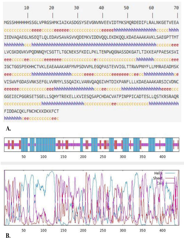
Secondary structure prediction of the GSRM chimeric protein using the GOR IV server: (A) predicted secondary structure composition (α-helix, extended strand, and random coil) and (B) graphical representation of the predicted secondary structure.
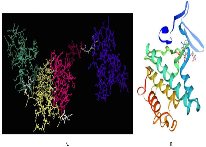
Predicted tertiary structure structure of the GSRM protein: (A) I-TASSER model showing domains (GRA1: blue, SAG1: pink, ROP18: yellow, MIC13: green, and linker: white) and (B) Three-dimensional structural model generated by the SWISS-MODEL server.
3.6. Tertiary Structure Validation
Ramachandran plot analysis using SWISS-MODEL showed that 84.90% of the residues were located in the favored regions, 10.41% in the allowed regions, and 4.69% in the outlier regions (Fig. 3). When more than 90% of the residues are located in the favorable and allowed regions and less than 5% are in the outlier regions, the GSRM has acceptable stereochemical quality.
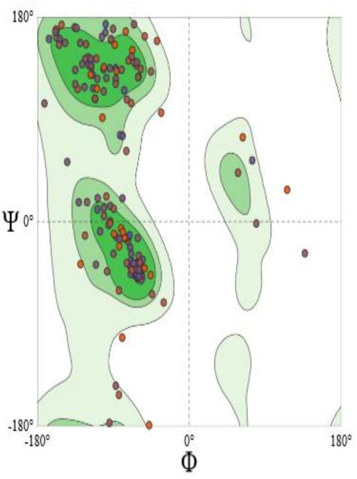
Validation of the predicted GSRM tertiary structure using Ramachandran plot analysis. The plot shows 84.90% of residues in favored regions, 10.41% in allowed regions, and 4.69% as outliers.
3.7. Antigenicity, Allergenicity, and Solubility Assessment
The VaxiJen score was estimated to be 0.6701. A common threshold of 0.5 is considered for potential protective antigens, and antigenicity above this threshold indicates strong antigenic potential. AlgPred analysis confirmed that the GSRM protein is nonallergenic. SOLpro predicted a solubility probability of 0.656861, and since values above 0.5 indicate that a protein is likely to be soluble when expressed in E. coli, this result suggests good expression potential in a prokaryotic system.
3.8. Prediction of Physicochemical Properties
The protein has a MW of 47,953.46 Da and a theoretical pI of 5.61. Negatively charged residues (Asp + Glu) total 61, and positively charged residues (Arg + Lys) total 49. Predicted half-life: 30 hours in mammalian reticulocytes (in vitro), > 20 hours in yeast (in vivo), and > 10 hours in E. coli (in vivo). The instability index is 43.07, and since values < 40 indicate stable proteins, this protein is considered unstable. The aliphatic index of the protein is 80.11, indicating good thermal stability. The GRAVY score of -0.316 indicates hydrophilicity.
3.9. Optimization of the Codon
Reverse translation and codon adaptation of the designed GSRM protein were performed using the EMBOSS Backtranambig tool on the EBI server, with E. coli as the host organism. This optimization replaces rare codons with synonymous codons preferred by E. coli, enhancing translational efficiency and minimizing ribosomal stalling.
3.10. mRNA Secondary Structure
The best-predicted structure had ΔG = −387.70 kcal/mol, and the first 10 nucleotides at the 5′ end did not form a stable hairpin or pseudoknot, supporting efficient ribosome binding and translation initiation (Fig. 4). These results indicate that the optimized GSRM transcript is suitable for robust expression in a prokaryotic system.
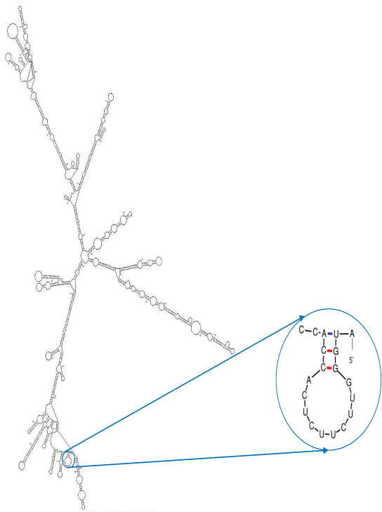
mFold-predicted secondary structure of the optimized GSRM sequence (ΔG = −387.70 kcal/mol).
4. DISCUSSION
The ability of T. gondii to evade the host immune response, its complex life cycle, and its antigenic diversity remain a challenge in the development of vaccines against this parasite. Therefore, this study aimed to develop a chimeric protein vaccine containing B- and T-cell epitopes of GRA1, SAG1, ROP18, and MIC13 antigens of T. gondii and analyze various aspects of the chimeric protein using different bioinformatics tools.
In this study, a variety of antigens (surface antigens such as SAGs and secretory antigens such as GRAs, ROPs, and MICs) were used for vaccine design. GRA1, SAG1, ROP18, and MIC13 were selected based on their roles in parasite survival, invasion, and pathogenicity. In addition, the aim was to select antigens that are expressed in all three stages of the parasite life cycle to elicit a more comprehensive immune response, as stage-specific antigens typically provide limited protection. On the one hand, combining epitopes from multiple antigens may elicit a broader and more effective immune response than a vaccine based on a single antigen [11, 14, 24]. This strategy is supported by recent immunoinformatics studies. For example, Nayeri et al. (2025) developed a novel chimeric vaccine candidate against T. gondii and demonstrated the feasibility of integrating epitopes from multiple antigens into a single construct with favorable predicted immunogenic and physicochemical properties [25]. Ghaffari and Rahimi (2024) constructed a multi-epitope peptide vaccine based on calcium-dependent protein kinases using immunoinformatics; their analyses suggested favorable antigenicity, solubility, and safety. Therefore, it becomes necessary to conduct experimental tests [26]. Similarly, another recent in silico study used tachyzoite-specific SAG1-related sequence proteins to design a peptide vaccine containing T-cell and B-cell epitopes that showed high antigenicity, non-allergenicity, and predicted interaction with human Toll-like receptor 4 [27]. Moreover, a genome-wide epitope vaccine study by Li et al. (2022) evaluated T. gondii membrane proteins and identified candidate epitopes for broad coverage, underscoring the power of comprehensive bioinformatics strategies [28]. In contrast, the present study introduces several methodological innovations. Rather than stitching together minimal epitopes, we selected antigenic fragments of approximately 100 amino acids from four antigens (GRA1, SAG1, ROP18, and MIC13), generated all 24 possible permutations of these fragments using the rigid A(EAAAK)A linker, and carefully screened the resulting structures for stability, folding quality, antigenicity, Ramachandran analysis, and physicochemical profile. This vaccine design strategy preserves the structural elements of the original antigens and could create a more stable and immunogenic vaccine candidate.
The selection of antigenic fragments was performed using rigorous epitope prediction analyses targeting B and T cell epitopes, ensuring that the designed construct could effectively stimulate an immune response. This is particularly important given the intracellular nature of T. gondii and the need for strong cellular immunity alongside humoral immunity. Notably, the A(EAAAK)A linker sequence used between the antigenic fragments has previously been shown to promote proper folding and functional independence of the individual domains, minimizing potential steric hindrance or epitope masking [29]. These design considerations are essential to ensure that the epitopes remain structurally accessible and immunologically active.
Our structural predictions showed that the GSRM protein has a balanced secondary structure, with a significant proportion of alpha-helices and random coils, which are favorable for stable folding and antigen presentation. Tertiary structure modeling revealed a moderate C-score (-2.21), indicating a relatively reliable three-dimensional conformation. Ramachandran plots confirmed the tertiary structure of the protein. The results showed that more than 90% of the residues were located in the favorable and allowed regions.
Moreover, immunoinformatic analyses predicted that the GSRM is antigenic (VaxiJen score: 0.6701) and nonallergenic, addressing two primary prerequisites for a safe and effective vaccine candidate. The pI is also one of the important physicochemical parameters that can be used to estimate the solubility of a protein at a certain pH, meaning that the protein precipitates in a solution close to the pI [24]. The pI is useful for establishing a suitable buffering system for protein purification based on pH-focused methods [24]. The presence of an acidic isoelectric point and a negative GRAVY score (-0.316) further indicates that the protein is likely to be hydrophilic and accessible to the host immune system, which is advantageous for immunogenicity. Although the instability index slightly exceeded the threshold for stability, the predicted solubility and favorable antigenicity scores suggest the protein remains a viable candidate for vaccine development. A MW (47953.46 Da) indicates that GSRM could be a suitable antigen (it has been indicated that antigens with a MW of < 5–15 kDa do not provide good immunity) [18] and an aliphatic index (80.11) indicative of good thermostability.
Codon optimization and mRNA secondary structure analysis are important steps in establishing a link between in silico vaccine design and practical protein production. High-throughput expression of antigen in E. coli facilitates purification and experimental testing. Codon optimization reduces the likelihood of ribosomal stalling, and stable mRNA folding, and the absence of inhibitory structures at the 5' end ensures translation of the chimeric protein.
This study is based exclusively on in silico analyses, which, although powerful for screening candidates, cannot fully simulate the complexity of immune responses in living systems. Structural predictions, antigenicity scores, allergenicity assessments, and solubility estimates are computational approximations that require experimental validation. Furthermore, epitope prediction tools vary in accuracy, and the selected fragments may behave differently when expressed as a recombinant protein in vitro or in vivo. The instability index suggests that the final construct may require stabilization strategies during expression and purification. Additionally, the immunogenic performance of the chimeric protein cannot be inferred without testing in animal models, including evaluation of cellular immunity, antibody responses, and assessing survival rates in mice infected with different strains. Therefore, the findings here should be considered preliminary and require experimental testing.
CONCLUSION
In conclusion, the bioinformatics-driven design of the GSRM protein represents a promising step towards the development of an effective multiepitope vaccine against T. gondii. By combining multiple antigens from different stages of the parasite's life cycle, this construct aims to overcome the limitations of single-antigen vaccines and provide broad, cross-stage protection. This work will provide a solid foundation for further experimental studies and help control toxoplasmosis in humans and animals.
AUTHORS' CONTRIBUTIONS
The authors confirm their contributions to the paper as follows: T.N.: conceived the study and designed the study protocol; T.N.: Was the supervisor of this research; E.N.: Performed the bioinformatics analysis; T.N.: Drafted the manuscript; E.N., M.F. and E.G.: Critically revised the manuscript; and All authors read and approved the final version of the manuscript.
LIST OF ABBREVIATIONS
| SAGs | = Surface antigens |
| MICs | = Microneme proteins |
| ROPs | = Rhoptry proteins |
| GRAs | = Dense granule proteins |
| NCBI | = National Center for Biotechnology Information |
| UniProt | = Universal protein resource |
| IEDB | = Immune Epitope Database |
| MHC | = Major histocompatibility complex |
| GOR IV | = Garnier-Osguthorpe-Robson IV |
| MW | = Molecular weight |
| pI | = Isoelectric point |
| GRAVY | = Grand average of hydropathicity |
| EBI | = European Bioinformatics Institute |
| MFE | = Minimum free energy |
| C-score | = Confidence score |
ETHICS APPROVAL AND CONSENT TO PARTICIPATE
This study did not involve human participants, animals, patient data, or biological specimens. However, the research project was registered and approved by the institutional ethics committee in accordance with national regulations governing research projects in medical universities. Therefore, an ethics approval code was assigned to the study (Ethics Code: IR.DUMS.REC. 1404.014). The ethics approval was administrative and regulatory in nature and did not reflect the involvement of human or animal subjects.
HUMAN AND ANIMAL RIGHTS
No animals/humans were used for studies that are the basis of this research.
AVAILABILITY OF DATA AND MATERIALS
The data and supportive information is available within the article.
ACKNOWLEDGEMENTS
The authors would like to thank the Infectious and Tropical Diseases Research Center, Dezful University of Medical Sciences, Dezful, Iran, for their support, cooperation, and assistance throughout the period of the study (IN&TR-404007-1404).