All published articles of this journal are available on ScienceDirect.

Molecular Typing and Occurrence of Extended-Spectrum Beta-Lactamases (ESBLs) Genes among Klebsiella oxytoca Isolated from Selected Hospitals in Khorramabad, Iran

Abstract
Introduction
Resistance of Klebsiella spp to various antibiotic families, such as beta-lactams, has increased due to the acquisition of plasmids carrying genes for Extended-Spectrum Beta-Lactamases (ESBLs). Today, ESBLs have become a major problem in healthcare settings.
Objective
This study aimed to determine the molecular fingerprinting and frequency of blaTEM, blaSHV,blaCTX-M, blaGES, blaPER, and blaVEB genes among Klebsiella oxytoca from clinical samples collected in selected hospitals in Khorramabad, Iran.
Methods
The present study was a cross-sectional study of Klebsiella oxytoca isolated from clinical specimens collected at selected hospitals in Khorramabad in 2019. After phenotypic identification of the isolates studied, antibiotic susceptibility patterns and beta-lactamase screening were performed using the disk diffusion method; genes encoding resistance were detected by PCR, and molecular fingerprinting was performed by PFGE.
Results
In this study, 32 K. oxytoca were isolated. The highest resistance rates were observed with ampicillin (93.8%) and cefotaxime (65.6%), and the lowest resistance rates were observed with colistin (0%), imipenem (15.6%), and amikacin (18.8%). 56.6% of isolates were ESBL- producing. Nineteen (59.4%), 2 (6.3%), and 16 (50%) isolates carried the blaTEM, blaSHV, and blaCTX-M genes, respectively. In addition, blaGES, blaPER, and blaVEB genes were not found in any of the tested isolates. Molecular typing results by the PFGE method showed that the isolates were very heterogeneous, and the 32 tested isolates were clustered in 18 pulse types.
Discussion
In the present study, K. oxytoca isolates exhibited concerning resistance to certain antibiotics, particularly cephalosporins. The high prevalence of ESBLs and blaTEM and blaCTX-M genes highlights the importance of continuous monitoring of these factors. On the other hand, the absence of blaGES, blaPER, and blaVEB genes and the high genetic heterogeneity of bacterial strains in PFGE indicate high strain diversity and the possibility of multiple sources of contamination.
Conclusion
Overall, the results of this study showed that the isolation and examination of K. oxytoca isolates in terms of diversity and molecular susceptibility profiling are important.
1. INTRODUCTION
Klebsiella oxytoca is a Gram-negative, nonmotile, encapsulated bacillus in the family Enterobacteriaceae. The genus Klebsiella generally includes two opportunistic pathogenic species, K. pneumoniae and K. oxytoca [1]. K. oxytoca is unique in nature and is isolated from skin, mucous membranes, oropharynx, and intestines of healthy humans and animals [1, 2]. Human infections caused by K. oxytoca are often associated with contamination from disinfectants, or parenteral fluid vials, humidifiers, and ventilators. The bacterium can be transmitted person-to-person, patient-to-patient, and staff-to-patient [3, 4].
First-line drugs for the treatment of Klebsiella spp infections are mainly cephalosporins [5]. However, in case of resistance to cephalosporins, aminoglycosides such as gentamicin, fluoroquinolones such as ciprofloxacin, or carbapenems are used [3]. Today, reports of resistance to various antibiotic families, including beta-lactams [6, 7], are emerging. Klebsiella spp has become resistant to various antibiotics, including extended-spectrum β-lactams, due to the acquisition of plasmids carrying genes for extended-spectrum β-lactamases (ESBLs). In Klebsiella spp., ESBLs can hydrolyze extended-spectrum cephalosporins, monobactams, and penicillins [8]. The distribution of β-lactamases, which are often carried by plasmids, is widespread worldwide; these plasmids also carry genes for resistance to aminoglycosides, sulfonamides, and other antibiotics [9]. In recent decades, ESBLs have become a serious problem in healthcare systems. Although the first β-lactam-resistant isolates were observed in hospital-acquired infections, they have now also spread in communities. In different geographical areas, including cities across Iran, ESBLs in the TEM, SHV, and CTX-M families have been identified. According to the results of studies conducted in Europe, the frequency of beta-lactamase-positive isolates is also higher than that of methicillin-resistant staphylococci [10, 11]. TEM, SHV, and CTX-M genes are widespread in Gram-negative bacilli of the Enterobacteriaceae family, especially in Escherichia coli and K. pneumoniae [12, 13]. The frequency of β-lactamase-producing isolates among Gram-negative bacilli in Asia is 39%, and the global average has been reported to be 16.3% [14-16]. Enterobacteriaceae strains isolated from healthcare facilities were producers of extended-spectrum β-lactamases. Epidemiological data from various parts of the world, including Africa, Asia, and the Middle East, have reported a high frequency of ESBL-positive isolates in clinical Klebsiella spp. isolates. In some studies, 50–70% of K. oxytoca isolates were reported to be ESBL producers. Such reports indicate a significant expansion of ESBL-carrying isolates, and the evaluation and detection of ESBL-carrying K. oxytoca strains could be important for infection control [17]. Given that β-lactamase enzymes can confer resistance to a wide range of cephalosporins, the presence of these enzymes delays the treatment of infections caused by β-lactam-resistant bacteria [18]. On the other hand, in epidemiological studies, molecular typing methods such as Pulsed-Field Gel Electrophoresis (PFGE) have received great attention because, in addition to determining the source of infections, they could be used to detect genetic diversity in bacteria, including changes in antibiotic resistance genes [19]. Therefore, the present study aimed to perform molecular fingerprinting and determine the occurrence of blaTEM, blaSHV, blaCTX-M, blaGES, blaPER, and blaVEB genes among K. oxytoca isolated from clinical samples from selected hospitals in Khorramabad city in 2019.
2. METHODS
2.1. Sample Collection
The present study was cross-sectional. The study population included all K. oxytoca isolates collected from clinical samples at Shahid Rahimi and Shohadaye Ashayer hospitals in Khorramabad, Iran, in 2019, as part of a census. Given that K. oxytoca isolates were received from the laboratory without the patient's name and with a code, there was no need for written consent from the patients.
Information on the samples, including patient age and gender, hospitalization department, and sample type, was recorded. Isolates were collected from various clinical specimens, including urine, blood, wounds, secretions, and body fluids. After culturing the samples on blood agar and MacConkey agar media, the colonies were diagnosed by biochemical and microbial tests, including Gram staining, catalase, oxidase, citrate consumption, motility, indole production, MR, VP, and sugar consumption in TSI medium [20].
2.2. Inclusion Criteria
The inclusion criteria are as follows:
- All clinical samples collected in 2019 from patients hospitalized or referred to Shahid Rahimi and Shohadaye Ashayer hospitals in Khorramabad city.
- Isolates that grew on Blood agar and MacConkey agar after cultivation.
- Samples that were identified as K. oxytoca based on biochemical and microbiological tests, including Gram stain, catalase, oxidase, citrate consumption, motility, indole production, MR and VP tests, and sugar fermentation in TSI medium.
- Clinical samples, including urine, blood, wound, secretions, and other body fluids.
- Samples for which the patients' demographic information (age, gender) and the relevant hospitalization ward were recorded.
2.4. Determination of Antibiotic Resistance Patterns and Detection of Extended-spectrum Beta-lactamase (ESBL)-producing Isolates
Antibiotic susceptibility pattern was determined by disk diffusion on Mueller-Hinton Agar (Merck, Germany) according to CLSI standard guidelines 2020 [21]. Ceftazidime, cefotaxime, ciprofloxacin, cefepime, aztreonam, imipenem, amikacin, gentamicin, nalidixic acid, and co-trimoxazole disks (Rosco, Denmark) were used in this study. To screen for ESBL-producing K. oxytoca, a half-McFarland bacterial suspension was cultured on Mueller-Hinton Agar. Then, 3 disks of ceftazidime, cefotaxime, and cefpodoxime alone and 3 discs in combination with clavulanic acid (ceftazidime/clavulanic acid, cefotaxime/clavulanic acid, and cefpodoxime/clavulanic acid) were placed on the culture medium. After incubation for 18-24 hours at 37°C, the diameters of the growth inhibition zones were measured. If the difference in the diameter of the zone of inhibition around at least one of the combined disks (containing clavulanic acid) compared to the single disk (without clavulanic acid) was more than 5 mm, the isolate was ESBLs-positive phenotype [22].
2.5. DNA Extraction and PCR
The SinnaPure DNA kit (SinaClon, Iran) was used to extract bacterial DNA (chromosomal and plasmid). All steps of DNA extraction were performed according to the kit instructions. In this study, the multiplex PCR method was used to detect the blaTEM and blaCTX-M genes. However, other genes were examined by simple PCR. The primer sequences and amplification temperature programs for the studied genes are shown in Tables 1 and 2, respectively. The PCR products were electrophoresed on 1% agarose, and the gels were finally visualized using a Gel Doc (BioDoc Analyse, Biometra). Pesedomonas aeroginosa strain containing blaGES, blaPER, blaVEB, and K. pneumoniae ATCC 7881 were used as positive gene controls.
| Primers | Sequence of Primers | Size of Amplicon (bp) | References |
|---|---|---|---|
| blaTEM | 5/-TCGCCGCATACACTATTCTCAGAATGA-3/ 5/-ACGCTCACCGGCTCCAGATTTAT-3/ |
445 | [23] |
| blaSHV | 5/-TTAGCGTTCCCAGTGCTC-3/ 5/-GGTTATGCGTTATATTCGCC-3/ |
865 | [24] |
| blaCTX-m | 5/-ATGTGCAGYACCAGTAARGTKATGGC-3/ 5/-TGGGT RAA RTA RGTSACCAGAA YCAGCGG-3/ |
593 | [24] |
| blaPER | 5/-GCTCCGATAATGAAAGCGT-3/ 5/-TTCGGCTTGACTCGGCTGA-3/ |
520 | [25] |
| blaVEB | 5/-CATTTCCCGATGCAAAGCGT-3/ 5/-CGAAGTTTCTTTGGACTCTG-3/ |
648 | [26] |
| blaGES | 5/-AGTCGGCTAGACCGGAAAG-3/ 5/-TTTGTCCGTGCTCAGGAT-3/ |
399 | [26] |
Note: *K is G or T, R is A or G, S is G or C, Y is C or T.
| blaTEM, blaCTX-M (Multiplex) | blaSHV | blaPER | blaGES | blaVEB | ||||||
|---|---|---|---|---|---|---|---|---|---|---|
| Temp | Time | Temp | Time | Temp | Time | Temp | Time | Temp | Time | |
| Initial denaturation | 94Cº | 10 min | 94Cº | 10 min | 94Cº | 10 min | 94Cº | 10 min | 94Cº | 10 min |
| Denaturation | 94Cº | 40 s | 94Cº | 1 min | 94Cº | 40 s | 94Cº | 40 s | 94Cº | 40 s |
| Annealing | 60Cº | 40 s | 58Cº | 1 min | 60Cº | 40 s | 60Cº | 40 s | 60Cº | 40 s |
| Extension | 72Cº | 1 min | 72Cº | 1 min | 72Cº | 1 min | 72Cº | 1 min | 72Cº | 1 min |
| No. of cycles | 30 cycles | 32 cycles | 30 cycles | 30 cycles | 30 cycles | |||||
| Final extension | 72Cº | 7 min | 72Cº | 10 min. | 72Cº | 7 min | 72Cº | 7 min | 72Cº | 5 min |
2.6. Pulsed Field Gel Electrophoresis (PFGE)
All 32 K. oxytoca isolates were typed using the PFGE technique according to the previously described procedure by Elahi et al. [27]. Briefly, the genomic DNA of the tested isolates and Salmonella enterica serovar Braenderup H9812 (as a DNA marker) was embedded in low-melting-point agarose (Thermo Scientific, Lithuania). Digestion was carried out with 20 U XbaI enzyme (Fermentas, Lithuania). The fragments were electrophoresed on a CHEF MAPPER apparatus (Bio-Rad, USA) for 22 h at 14°C, with a switch time of 5-35 s, a voltage gradient of 120°, and a field strength of 6 V/cm. Gels were stained with ethidium bromide and visualized by the UVItec Gel Doc system (UVItec, UK). The obtained electrophoretic patterns were compared using BioNumerics software, version 7.1 (Applied Maths, Sint-Martens-Latem, Belgium). The Dice coefficient calculated the similarities, and the unweighted pair-group method was used for cluster analysis (UPGMA). The similarity of more than 80% between the DNA patterns of isolates was considered a clone.
3. RESULTS
In this study, 32 isolates of K. oxytoca were identified using biochemical tests. The isolates were Gram-negative coccobacilli, citrate-positive, indole-positive, VP-positive, catalase-negative, MR-negative, and negative for acid production, glucose and lactose fermentation, and H2S production in TSI medium. Of the 32 K. oxytoca isolates, 21 (65.6%) were isolated from women and 11 (34.4%) from men. The majority of subjects belonged to the 41–60 age group (10 subjects, 31.3%). The mean age of the subjects whose samples were included in the study was 46.16 ± 22.18 years. The youngest subject was 10 years old, and the oldest was 84 years old. Most isolates were from urine samples (28, 87.5%), followed by wounds and sputum (2 each, 6.3%). Of the total samples, 28.1% were collected from Shahid Rahimi Hospital and 71.9% from Shohadaye Ashayer Hospital. In addition, most of the samples were from the outpatient ward, accounting for 53.1% (Table 3).
| Variable | Category | Frequency (%) |
|---|---|---|
| Gender | Male | 11 (34.4) |
| Female | 21 (65.6) | |
| Age groups | < 20 | 6 (18.8) |
| 21–40 | 7 (21.9) | |
| 41–60 | 10 (31.3) | |
| 61–80 | 7 (21.9) | |
| > 80 | 2 (6.3) | |
| Source of samples | Urine | 28 (87.5) |
| Wound | 2 (6.3) | |
| Sputum | 2 (6.3) | |
| Hospitals of sampling | Shohadaye Ashayer | 23 (71.9) |
| Shahid Rahimi | 9 (28.1) | |
| Sampling wards | Outpatient | 17 (53.1) |
| Women | 4 (12.5) | |
| Urology | 2 (6.3) | |
| ICU | 4 (12.5) | |
| Surgery | 2 (6.3) | |
| Burn | 3 (9.4) |
3.1. Antibiotic Susceptibility Pattern
Among the 32 K. oxytoca isolates collected, the resistance rate to antibiotics ceftazidime, cefotaxime, ciprofloxacin, cefepime, aztreonam, imipenem, amikacin, gentamicin, nalidixic acid, co-trimoxazole and colistin was (43.8%) 14, (65.6%) 21, (46.9%) 15, (31.3%) 10, (31.3%) 10, (15.6%) 5, (18.8%) 6, (31.3%) 10, (59.4%) 19, (59.4%) 19 and (0.0%) 0, respectively. Therefore, the highest resistance rate was to ampicillin and cefotaxime, and the lowest was to colistin, imipenem, and amikacin.
3.2. Screening for ESBL-producing K. oxytoca
Among 32 K. oxytoca strains, 21 (56.6%) were phenotypically ESBL-producing. The distribution of ESBL-positive isolates according to the source of specimens, patients’ gender, ward, and hospital is shown in Table 4. There was no significant relationship between the variables studied and the frequency of ESBL-producing K. oxytoca isolates (p<0.05).
| Variable | Category | Positive n (%) | Negative n (%) | Chi-square (X2) | p-value |
|---|---|---|---|---|---|
| Age groups | < 20 | 4 (66.7) | 2 (33.3) | 1.582 | 0.850 |
| 21–40 | 4 (57.1) | 3 (42.9) | |||
| 41–60 | 8 (80.0) | 2 (20.0) | |||
| 61–80 | 4 (57.1) | 3 (42.9) | |||
| > 80 | 1 (50.0) | 1 (50.0) | |||
| Source of samples | Urine | 19 (67.9) | 9 (32.1) | 0.415 | 0.998 |
| Wound | 1 (50.0) | 1 (50.0) | |||
| Sputum | 1 (50.0) | 1 (50.0) | |||
| Hospitals of sampling | Shohadaye Ashayer | 14 (60.9) | 9 (39.1) | 0.82 | 0.441 |
| Shahid Rahimi | 7 (77.8) | 2 (22.2) | |||
| Gender | Male | 7 (63.6) | 4 (36.4) | 0.029 | 0.998 |
| Female | 14 (66.7) | 7 (33.3) | |||
| Sampling wards | Outpatient | 10 (58.8) | 7 (41.2) | 4.142 | 0.605 |
| Women | 3 (75.0) | 1 (25.0) | |||
| Urology | 2 (100.0) | 0 | |||
| ICU | 3 (75.0) | 1 (25.0) | |||
| Surgery | 2 (100.0) | – | |||
| Burn | 1 (33.3) | 2 (66.7) |
3.3. The Distribution of blaTEM, blaSHV, blaCTX-M, blaGES, blaPER, and blaVEB genes
Based on the results of PCR tests, the frequency of blaTEM, blaSHV, and blaCTX-M genes was 19 (59.4%), 16 (50%), and 2 (6.3%). Also, the frequency of blaGES, blaPER, and blaVEB genes was zero. As shown in Table 5, the association between gender and the frequency of blaCTX-M genes was statistically significant (p=0.012). In addition, the association between hospitals and the presence of blaSHV (p=0.02) and blaCTX-M (p=0.049) genes was statistically significant. Conversely, the difference in the distribution of blaTEM, blaSHV, and blaCTX-M genes by source of samples, wards, and age groups was not statistically significant.
| blaCTX-M | blaTEM | blaSHV | ||||||
|---|---|---|---|---|---|---|---|---|
| Variable | Category | Positive n (%) | Negative n (%) | Positive n (%) | Negative n (%) |
Total n (%) |
Positive n (%) | Negative n (%) |
| Source of samples | Urine | 15 (53.6) | 13 (46.4) | 16 (57.1) | 12 (42.9) | 28 (100) | 2 (7.1) | 26 (92.9) |
| Wound | – | 2 (100) | 1 (50.0) | 1 (50.0) | 2 (100) | – | 2 (100) | |
| Sputum | 1 (50.0) | 1 (50.0) | 2 (100) | – | 2 (100) | – | 2 (100) | |
| Chi-square (X2) | 2.143 | 1.499 | 0.305 | |||||
| P-value | 0.731 | 0.757 | 0.999 | |||||
| Sampling wards | Outpatient | 8 (47.1) | 9 (52.9) | 9 (52.9) | 8 (47.1) | 17 (100) | 1 (5.9) | 16 (94.1) |
| Women | 4 (100) | – | 3 (75.0) | 1 (25.0) | 4 (100) | – | 4 (100) | |
| Urology | – | 1 (100) | 2 (100) | – | 2 (100) | – | 2 (100) | |
| ICU | 2 (50.0) | 2 (50.0) | 2 (50.0) | 2 (50.0) | 4 (100) | 1 (25.0) | 3 (75.0) | |
| Surgery | 1 (50.0) | 1 (50.0) | 1 (50.0) | 1 (50.0) | 2 (100) | – | 2 (100) | |
| Burn | 1 (33.3) | 2 (66.7) | 2 (66.7) | 1 (33.3) | 3 (100) | – | 3 (100) | |
| Chi-square (X2) | 5.365 | 2.35 | 3.137 | |||||
| P-value | 0.449 | 0.873 | 0.721 | |||||
| Hospitals of sampling | Shohadaye Ashayer | 9 (39.1) | 14 (60.9) | 13 (56.5) | 10 (43.5) | 23 (100) | – | 23 (100) |
| Shahid Rahimi | 7 (77.8) | 2 (22.2) | 6 (66.7) | 3 (33.3) | 9 (100) | 2 (22.2) | 7 (77.8) | |
| Chi-square (X2) | 3.865 | 0.276 | 5.452 | |||||
| P-value | 0.049 | 0.704 | 0.020 | |||||
| Gender | Male | 2 (18.2) | 9 (81.8) | 6 (54.5) | 5 (45.5) | 11 (100) | – | 11 (100) |
| Female | 14 (66.7) | 7 (33.3) | 13 (61.9) | 8 (38.1) | 21 (100) | 2 (9.5) | 19 (90.5) | |
| Chi-square (X2) | 6.788 | 0.162 | 1.117 | |||||
| P-value | 0.012 | 0.721 | 0.534 | |||||
| Age groups | < 20 | 4 (66.7) | 2 (33.3) | 3 (50.0) | 3 (50.0) | 6 (100) | 1 (16.7) | 5 (83.3) |
| 21–40 | 2 (28.6) | 5 (71.4) | 6 (85.7) | 1 (14.3) | 7 (100) | – | 7 (100) | |
| 41–60 | 6 (60.0) | 4 (40.0) | 5 (50.0) | 5 (50.0) | 10 (100) | 1 (10.0) | 9 (90.0) | |
| > 60 | 4 (44.4) | 5 (55.6) | 5 (55.6) | 4 (44.4) | 9 (100) | – | 9 (100) | |
| Chi-square (X2) | 2.463 | 2.657 | 2.418 | |||||
| P-value | 0.505 | 0.483 | 0.550 | |||||
3.4. Molecular typing by PFGE
PFGE analysis was successfully performed on all 32 K. oxytoca isolates. To define clusters (pulse types), isolates with at least 80% similarity in PFGE patterns were grouped into a cluster, in accordance with standard PFGE protocols and previous studies. Based on this criterion, the isolates were divided into 18 distinct clusters, indicating high genetic diversity (Fig. 1). The largest cluster (cluster no. 10) contained 7 isolates (21.9%; 7 out of 32). Epidemiological investigation showed that all isolates in this cluster were collected from the outpatient ward, and no strong epidemiological links were observed among them. The other 25 isolates (78.1%; 25 of 32) were grouped into 17 clusters with one to three members, further confirming that there is significant genetic diversity among isolates and no clear clonal expansion is observed.
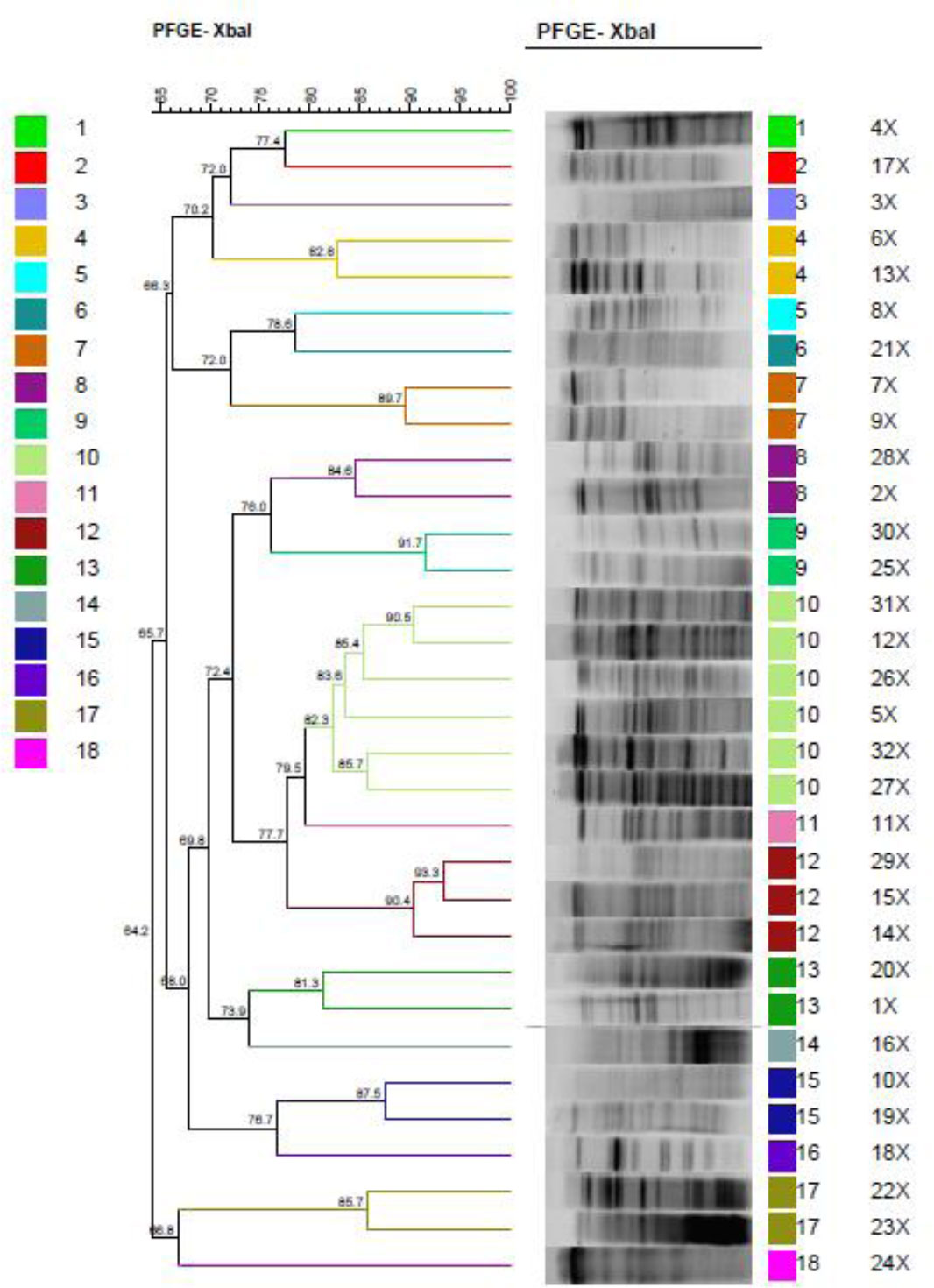
PFGE typing of the tested K. oxytoca isolates. Electropherogram of PCR products from 52 tested samples, digested with the Xba1 enzyme. Isolates were divided into 18 distinct clusters. The largest cluster (cluster no. 10) contained 7 isolates collected from the outpatient ward. The fragments were electrophoresed by the CHEF MAPPER apparatus (Bio-Rad, USA) for 22 h at 14°C. Gels were stained with ethidium bromide and visualized by the UVItec Gel Doc system (UVItec, UK). The electrophoretic patterns obtained were compared using BioNumerics software, version 7.1
4. DISCUSSION
K. oxytoca is a member of the Enterobacteriaceae family and is reported less frequently than K. pneumoniae. In the present study, 32 K. oxytoca isolates were obtained from various clinical specimens collected from wards of Shahid Rahimi and Shohadaye Ashayer Teaching Hospitals in Khorramabad over a 1-year period. The prevalence observed in this study is comparable to previous reports from Iran and other countries, including studies conducted in Tehran (1–12.8%), Shahrekord (2.2%), and Burkina Faso (14.06%), confirming the relatively low frequency of K. oxytoca compared with K. pneumoniae [28–31]. K. oxytoca is recognized as an important pathogen across all age groups. In the present study, the highest isolation rate was observed in patients aged 41–60 years. This finding differs from those reported by Validi et al., who found the highest prevalence in the 16–30-year age group [28], and by Khodaparast et al., who found the highest prevalence in children aged 1–15 years [32]. These discrepancies may be attributed to differences in patient populations, geographic regions, and specimen sources. Cephalosporins, carbapenems, and aminoglycosides are commonly used as first-line agents for the treatment of Klebsiella infections [33, 34]. However, increasing resistance to cephalosporins and carbapenems has become a major concern worldwide, largely due to the acquisition of plasmid-mediated resistance genes, particularly those encoding Extended-Spectrum Β-Lactamases (ESBLs) [35, 36]. In the present study, none of the isolates showed resistance to colistin. Similarly, Nassari et al. reported complete susceptibility of K. oxytoca isolates to imipenem and colistin in hospitals in Shiraz [37], findings that are in agreement with ours. In contrast, high resistance rates were observed against amikacin, cefoxitin, ciprofloxacin, and cefepime. These results indicate that many β-lactam antibiotics are no longer effective for treating infections caused by K. oxytoca in this region. Similar resistance patterns have been reported by Validi et al., who observed variable susceptibility of K. oxytoca isolates to gentamicin, carbapenems, cephalosporins, and aztreonam, with the highest resistance to ampicillin, amoxicillin, and ticarcillin [28]. Furthermore, studies from Iraq have reported high resistance rates to β-lactams and carbapenems among urinary isolates of K. oxytoca, emphasizing the risk of emerging multidrug resistance and the need for continuous surveillance [38]. The variability in antimicrobial resistance patterns across studies and regions reflects differences in antibiotic consumption, infection control practices, sample sources, and the timing of isolation collection. Even within the same geographic region, resistance profiles may change over time, highlighting the importance of periodic monitoring to guide appropriate empirical therapy [39]. In the present study, 56.6% of the isolates were identified as ESBL producers based on phenotypic methods, which is higher than rates reported in several previous studies, including those conducted in India (25%), the Netherlands (2.4%), and Nepal (16.7%) [40–42]. Molecular analysis revealed that blaTEM, blaCTX-M, and blaSHV genes were present in 59.4%, 50%, and 6.3% of isolates, respectively, while blaGES, blaPER, and blaVEB were not detected. These findings differ from studies conducted in Iran, Tunisia, Lebanon, and Egypt, where lower frequencies of ESBL genes among K. oxytoca isolates were reported [43–47]. The absence of blaPER in our isolates is in line with the findings of Nakhaei Moghaddam et al. in Tehran [48]. Recent studies from Iraq and Japan have reported a high burden of resistance genes, including blaCTX-M, blaAmpC, and blaSHV, in both human and animal K. oxytoca isolates, indicating the potential for widespread dissemination of resistance genes [49, 50]. Whole-genome sequencing studies from China have also demonstrated high genomic diversity among K.oxytoca complex isolates, supporting the heterogeneous nature of this species [51]. PFGE analysis in the present study revealed high genetic diversity, with 32 isolates grouped into 18 clusters at ≥80% similarity, suggesting multiple sources rather than clonal spread. This diversity may be related to patient referral from different healthcare centers within the province.
One of the main reasons for the heterogeneity among K. oxytoca strains in our study could be the diversity in sample collection, wards, and the inclusion of inpatients and outpatients. In addition, Shahid Rahimi and Shohada Ashayer hospitals are considered provincial referral hospitals, which leads to the admission of diverse patients from across the province, resulting in genetic diversity of strains.
The high prevalence of ESBL-producing K. oxytoca has significant clinical implications, including limited therapeutic options, increased reliance on carbapenems, prolonged hospitalization, higher healthcare costs, and increased mortality. Therefore, strict infection control measures, rational antibiotic use, and implementation of antimicrobial stewardship programs are essential. As the first study to investigate K. oxytoca isolates in Lorestan Province, this research provides valuable baseline data on antimicrobial resistance patterns and the distribution of ESBL genes. Further studies focusing on additional resistance mechanisms, including metallo-β-lactamases, are recommended to improve treatment strategies and limit the spread of antimicrobial resistance.
5. LIMITATIONS
The following are some of the limitations of this study:
A) The number of isolates studied was relatively limited, which may affect the generalizability of the results.
B) Samples were collected only from selected hospitals in one city; therefore, the resistance pattern in other regions or treatment centers may be different.
C) Due to financial and equipment limitations, it was not possible to examine a wider range of resistance genes or complementary molecular typing methods.
CONCLUSION
Given the increasing use of antibiotics in hospitals and the community, and the growing spread of ESBL-producing isolates, it is necessary to identify ESBL-producing isolates and determine their antibiotic resistance patterns before prescribing antibiotics. This will allow patients with serious infections to be prescribed beta-lactam antibiotics with the greatest possible efficiency.
AUTHORS’ CONTRIBUTIONS
The authors confirm their contributions to the paper as follows: G.H.G.: Conceived the study and supervised the project; P.S.H and G.H.G.: Curated the data and wrote the manuscript; H.À.: Performed the data analysis; T.Z.: Contributed to the content of the present manuscript and participated in revising the paper.
LIST OF ABBREVIATIONS
| K. oxytoca | = Klebsiella oxytoca |
| ESBLs | = Extended-spectrum beta-lactamases |
| PFGE | = Pulsed-field gel electrophoresis |
| CLSI | = Clinical and Laboratory Standards Institute |
ETHICS APPROVAL AND CONSENT TO PARTICIPATE
All procedures related to clinical samples were approved by the Ethics Committee of Lorestan University of Medical Sciences, Iran (IR.LUMS.REC.1400.166).
HUMAN AND ANIMAL RIGHTS
All human research procedures followed were in accordance with the ethical standards of the committee responsible for human experimentation (institutional and national), and with the Helsinki Declaration of 1975, as revised in 2013.
AVAILABILITY OF DATA AND MATERIALS
The data and supportive information are available within the article.
FUNDING
The funding for this research was provided by Lorestan University of Medical Sciences, Iran (IR.LUMS.REC.1400.166).
ACKNOWLEDGEMENTS
The authors hereby express their gratitude for the cooperation of the Vice-Chancellor for Research and Technology of Lorestan University of Medical Sciences, Iran.