All published articles of this journal are available on ScienceDirect.

Prevalence and Recovery of Microorganisms from Containers used for the Collection of Forensic Biological Samples
Authors Info & Affiliations
Abstract
Background:
Microbes play a significant role in the degradation of biological evidence collected for forensic analysis. The present study is aimed to isolate and identify the microbes present inside the empty container used for the biological evidence collection.
Methods:
Bacterial isolation from the selected containers was done by cotton swab over the inner surface of the containers. Streaking was done on the surface of the three different culture plates as a Blood agar plate, Nutrient plate and MacConkey plate. The plates were placed in an incubator shaker at 37ºC for 48 hours. The colonies grown on the surface of the media were counted on and used for further study. Various biochemical assays were performed to characterize isolated bacteria.
Results:
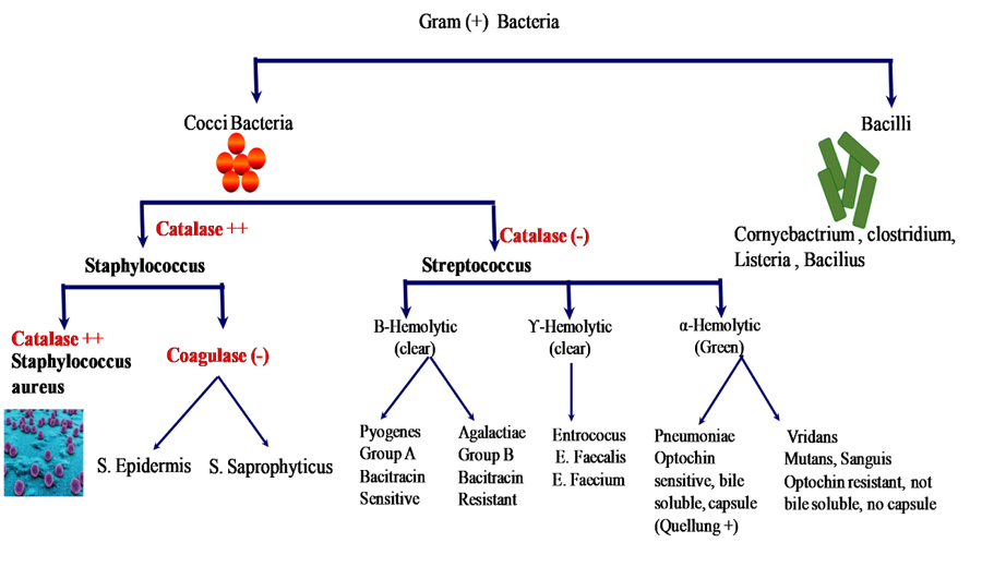
Staining results suggested that the presence of Gram-positive stain (Staphylococcus, Bacillus, Corynebacterium, Clostridium) and Gram negative stain (E. coli, Enterobacteriaceae, Pseudomonas, Salmonella, Shigella, Stenotrophomonas, Bdellovibrio, Acetic acid bacteria). The Catalase and Coagulase test suggested the presence of Staphylococcus aureus, S. epidermis and S. sapropyticus. Moreover, the indole test suggested the presence of Citrobacter koseri, Kebsiella oxytoca, Proteus vulgaris etc. Some of the bacteria were urea metabolizing, including Proteus spp, Helicobacter pylori, Cryptococcus spp, Corynebacterium spp.
Conclusion:
This study recommends that there should be proper maintenance of the chain of custody from the collection to analysis so that evidence properly prevents degradation or contamination in the biological evidence. Extra care is needed for the collection and packing of biological evidence from the crime scene. Moreover, the collection containers, if left wide open, lead to contamination and degradation of biological evidence.
1. INTRODUCTION
The collection of biological evidence at a crime scene is an essential step in solving medico-legal cases. Any error in sample collection, handling, and preservation, may affect forensic analysis that might interrupt the judgment process [1, 2]. High moisture content, bacterial growth or degradation rate of biological evidence often limit the easy retrieval and identification of genetic materials like RNA and DNA from the trace biological samples [3, 4]. As a result, biological evidence requires more caution while collecting, handling, transporting, and storing to obtain meaningful information from the samples collected [5-7]. Various factors, such as tissue type, collection period, containers, preservatives, and other additives used, shipment, transit period, directly affect the quality of the samples and the stability of biomarkers in the biological sample. Therefore, these points should be addressed, and due care should be taken during the evidence collection and transportation procedures [2, 7, 8]. Furthermore, the possibility of contamination becomes manifold due to inadvertent addition of DNA (exogenous), RNA, amino acids, and enzymes from other sources (DNA from analyst, other DNA samples in the laboratory, and Macrobacteria, protozoan) get mixed with the sample that to be analyzed [4, 9].
To introduce the DNA findings in the court of law, it becomes more significant that the evidence collection and preservation methods are given significant priority. The maintenance of the integrity of evidence commences with the arrival of the investigation officer at the crime scene. The evidence collection methods employed by the Investigating officer will also depend upon the state and condition in which the biological evidence is found at the scene of the crime. However, it is always advisable to collect relevant and sufficient quantity of evidence from the crime scene, although this aspect also can pose the danger of adding on additional dirt, grease, fluids, and other material from the surrounding area. Hence contaminated sample will be adversely affecting the DNA typing process. Standard Protocols are in place, which should be followed while collecting the biological specimen. These protocols ensure minimizing the specimen deterioration.
Use of inappropriate and non-sterile containers, contaminated instruments, wearing used gloves, cross-contamination with unknown samples can lead to inconsistent results, inaccurate data, and incorrect concentration measurements [10, 11], which in turn can affect judgment. Therefore investigators and laboratory personnel must be conscientious about contamination issues when identifying, collecting, and preserving samples [12]. Studies have shown that the isolation of high-quality DNA and the rate of human DNA degradation are exponentially linked with the growth rate of bacteria and other pathogens present in the sample. The growth of microbes depends on environmental factors like temperature, light, pH, osmotic conditions, nutrients and humidity [13]. Microorganisms are the most abundant biological entities of the biosphere which perform many essential functions; ranging from human health to environmental activities [14]. While performing diverse functions, these invisible communities might alter the forensically significant evidence as they may degrade DNA in the specimen [15]. With this background, the present study is focused on figuring out the presence of microbes in the biological sample collection containers and identifying and isolates these biological entities responsible for biological contamination and degradation of the forensically important specimens.
2. MATERIALS AND METHODS
i). Chemicals and Reagents
Culture media plates as a Blood Agar plate, Nutrient Agar plate, MacConkey Agar plate (LifeSciences) were used for microbial culture. The stains used were Crystal violet (C25N3H30Cl), Iodine and Safranin (C20H19ClN4). General chemicals used were Ethyl alcohol, Hydrogen peroxide (3%), Citrated plasma, Yeast extract, Potassium phosphate, Potassium Monobasic and Dibasic buffer, Urea, Phenol red, Lysis Buffer, Proteinase K.
ii). Instruments
Biosafety Cabinet Class II with MS powder Coated Stand Burn out Stand (Envision Biotech), Electric Loop Steriliser (York Scientific Industries), Bacteriological Incubator (Vijay Surgical Corporation), Microscope Lambomed Vision 2000 (Labomed), Thermo-mixer (Eppendorf), Vortex (Scientific Systems), Automated DNA Extraction System (Applied Biosystems), NanoDrop 2000 (Thermo Scientific).
2.1. Methodology
2.1.1. Selection of Containers
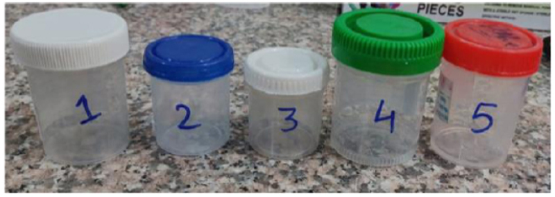
The sample containers were obtained from the Police station (Police Station: Sector 58, Noida, Uttar Pradesh, India) with proper consent. Five polystyrene containers were collected and analyzed for microbial study at the Shriram Singh Multispecialty Hospital, Delhi. The sample containers were selected based on their utility, availability, usage. It is significant to note that the type of containers may vary in the police stations and forensic laboratories. The obtained unsterilized containers were placed at room temperature for further study. The detailed descriptions of the containers are as given below (Fig. 1):
- Sample 1- 40ml Screw-capped Vials (White Cap)
- Sample 2- 20ml Screw-capped Vials (Blue Cap)
- Sample 3- 10ml Screw-capped Vials (White Cap)
- Sample 4- 40ml Screw-capped Vials (Green Cap)
- Sample 5- 30ml Screw-capped Vials (Red Cap)
2.1.2. Microbial Culture of Containers by Antonie Van Leeuwenhoek Method
2.1.2.1. Collection of Microbial Extract from Containers
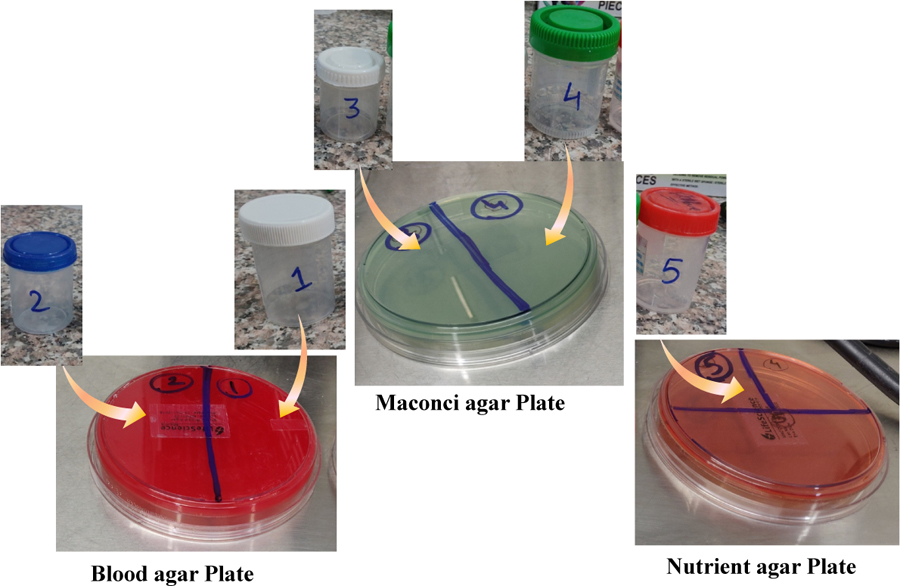
A sterile cotton swab was taken and thoroughly moved over the container’s inner surface. Streaking was done by cotton swab on the surface of the three different culture plates as Blood agar plate, Nutrient plate, MacConkey plate as shown in Fig. (2). After successful streaking, the plates were kept in an incubator shaker at 37ºC for 48 hours. The colonies grown on the surface of the media were then counted and used for further study. The bacterial culture was prepared according to the Lawn Culture (or carpet culture) Technique. Briefly, the bacterial culture was spread over an agar plate by using a sterile cotton swab. Alternatively, an automated dispenser machine was used to spread bacteria amount evenly on a plate that was achieved by rotation of plate [16].
2.3. Staining of Bacterial Colonies by Hans Christian Gram Method
For this procedure, a few of the colonies were picked up using a metallic inoculating loop. The smear of those colonies was made by adding a drop of distilled water on clean slides. Primary staining was done by crystal violet followed by iodine and then discoloration was done using alcohol. Finally, staining was done using Safranin (C20H19ClN4). These stained slides were then observed under a microscope at 100X objective magnification with oil immersion technique.
2.4. Assays for Biochemical Tests
2.4.1. Catalase Enzyme Assay
A small amount of colony growth smear was transferred on the surface of a sterile dry glass slide by using a sterilized inoculating loop. On adding 2-3 drops of 3% H2O2 solution over the glass slide with microbial colony smear, catalase producing microbes react with H2O2 and evolve oxygen bubbles [17].
2.4.2. Coagulase Test
This test is used to differentiate Staphylococcus aureus in the sample of interest. A thin bacterial smear emulsified with water was placed over a glass slide to form a concentrated film. Simultaneously, the positive and negative controls of the strains were performed to confirm the good reactivity of the plasma. Then a sterilized inoculating wire was dipped into the undiluted plasma solution at standard temperature and pressure (STP). Then adhering traces of plasma were stirred into the staphylococcal suspension on the slide. A similar procedure was also followed for the control suspensions as well [18].
2.4.3. Indole Test
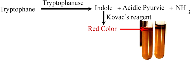
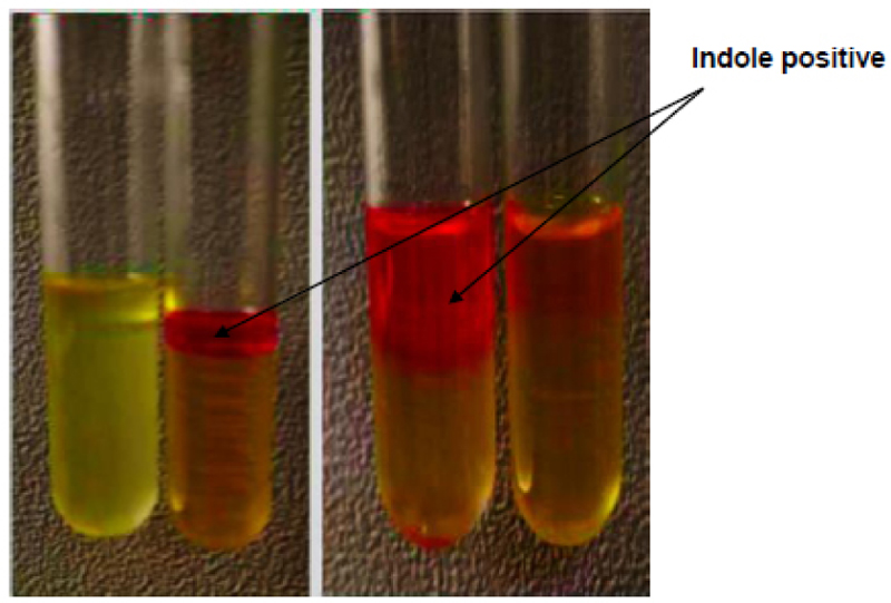
Briefly, inoculated L-tryptophan broth was taken as a substrate in the microorganism test [19, 20]. The mixture was then incubated at 35ºC for 48 hours at STP. After incubation, a mixture of 4-5 drops of Kovacs Reagent (Isoamyl alcohol + para-dimethylaminobenzaldehyde (DMAB), concentrated hydrochloric acid) was added to each tube and then vortexed gently. The substrate (L-Tryptophan) is converted into Indole and pyruvic acids with the release of ammonia in the presence of Tryptophanase enzyme. Kavoc reagent reacts with Indole to form a red-colored complex (para-dimethylaminobenzaldehyde) as shown in Fig. (3).
2.4.4. Urease Test
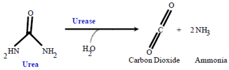
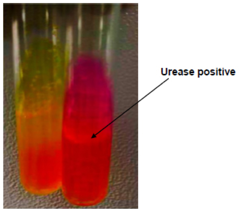
The Urease test is used for differentiating microorganism based on their ability to hydrolyze urea. The isolated bacterial colonies were grown on agar slants. To this one-two drops of an overnight brain-heart infusion broth culture were added. The reaction mixture was incubated and then isolated bacterial colonies were grown at 35±2°C in ambient air for 48 hours. The pink color development was examined for 3-7 days [21, 22] (Fig. 4).
2.5. DNA Isolation and Quantification
Isolation of bacteria DNA was done by Automated Extraction method procedure involve Lysis, Precipitation, and Purification. Briefly, sterile cotton buds were taken to pick up few randomly grown colonies. The whole portion of that cotton bud containing the colonies was added into the Eppendorf tubes. After that lysis buffer (50 µl) was added, followed by proteinase K (50 µl) and then magnetic beads and kept in the Automated DNA Extraction machine. Quantification of DNA samples was done by the Nanodrop 2000 spectrophotometer instrument (Thermo Scientific™) further data was analyzed by using Thermo Software IQ software. Briefly, 2 μL isolated DNA samples were added on the sample holder platform (Nanodrop) by sterile pipette and DNA quantification was done by selecting UV Visible Maxwell RSC software.
3. RESULTS
3.1. Micro Bacteria Culture Results
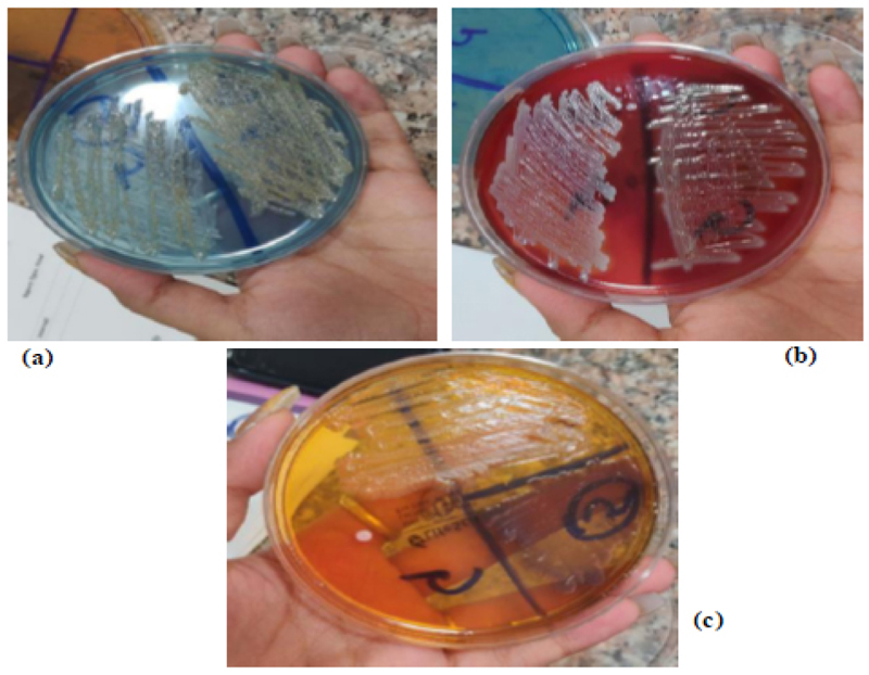
Earlier, the researcher used different agar methods to culture the microbes due to their compatible benefits. In this study, micro bacteria extracted from the containers were grown on all three selected types of culture plates as Blood Agar (Titan Biotech Blood Agar), MacConkey Agar (Titan Biotech Blood Agar), and Nutrient Agar (Titan Biotech Blood Agar) as the variety of three different plates showed different types of grown microorganism.
Moreover, the Nutrient agar is found to be non-selective, as it supports the growth of a broad range of bacteria like Bacillus subtilis, E.coli, Pseudomonas fluorescens as shown in (Fig. 5). In comparison, MacConkey agar inhibits the growth of Gram-negative bacteria like Bacillus subtilis, E Coli (selective medium), Pseudomonas fluorescens. Furthermore, MacConkey agar is also a differential medium that distinguishes between lactose metabolizing Gram-negative bacteria and those that do not metabolize. Blood agar plates (BAPs) contain 5-10% mammalian blood (usually sheep or horse) i.e. why used to grow a wide range of pathogenic bacteria such as Haemophilus influenzae, Streptococcus pneumoniae, and Neisseria species. Blood agar media plates act as differentiate media for the growth of certain strains of Bacillus, Enterococcus, Staphylococcus, and Aerococcus.
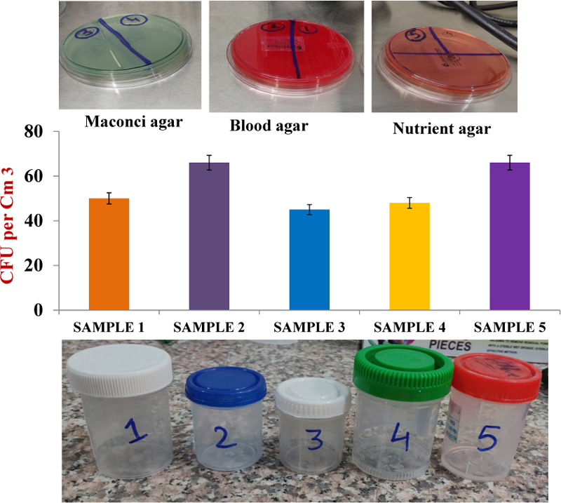
All the containers (05) used in the present study were found to be contaminated with microbes. The number of bacterial colonies are shown in Table 1:
| Samples | No. of Colonies |
|---|---|
| Sample 1 -40ml Screw-capped Vials (White Cap) | 55 |
| Sample 2 -20ml Screw-capped Vials (Blue Cap) | 65 |
| Sample 3 -10ml Screw-capped Vials (White Cap) | 45 |
| Sample 4 -40ml Screw-capped Vials (Green Cap) | 48 |
| Sample 5 -30ml Screw-capped Vials (Red Cap) | 63 |
In the colony counter study, the highest number of colonies was observed in sample-2 (65 CFU) on blood agar media followed by sample-5 (63CFU) on nutrient agar media, as shown in Fig. (6).
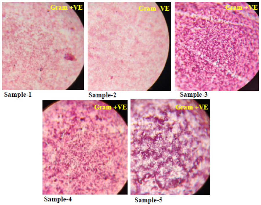
3.2. Bacterial Staining
Bacteria can be classified into Gram-positive and Gram-negative groups according to structural differences found on the cell wall. Certain bacteria retain more crystal violet and reflect violet color because of the thick layer of peptidoglycans on their cell. These are said to be Gram-positive bacteria. In contrast, Gram-negative which does not retain the crystal violet (due to thinner peptidoglycans wall) thus stain red and pinkish after destaining process as shown in Fig. (7). In this study, results showed that four slides were gram–positive suggesting the presence of Staphylococcus, Bacillus, Corynebacterium, clostridium. On other hand, Gram-negative stain which might suggest the presence of E. coli, Enterobacteriaceae, pseudomonas, Salmonella, Shigella, Stenotrophomonas, Bdellovibrio, Acetic acid bacteria as mentioned in Table 2.
3.3. Biochemical Results
The purpose of biochemical tests was to find their characteristics and presence in culture plates.
3.3.1. Catalase Slide Test
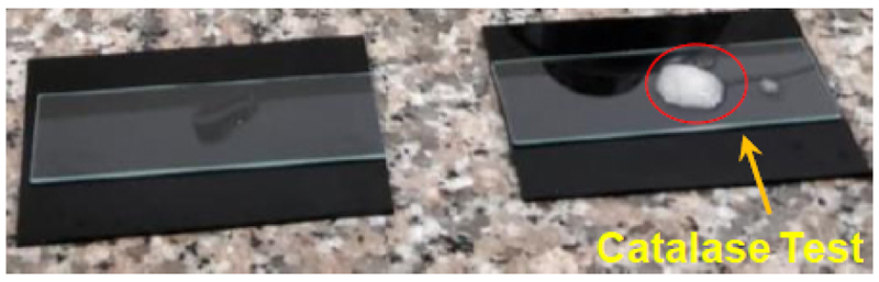
Catalase test was performed to identify organisms that produce the catalase enzyme (antioxidant). It differentiates catalase-producing microbacteria like staphylococci with non-catalase-producing microbacteria like streptococci. An enzyme catalase is produced by aerobic bacteria that protect the cell from the toxic by-products of oxygen metabolism. One minute after adding the hydrogen peroxide, clumping in Catalase reagent was observed with slight effervescence (bubble formation). This signifies the catalase-positive test, as shown in Fig. (8). Catalase-negative bacteria incapable of respiring using oxygen (like Streptococci) i.e why said to be as anaerobes or facultative anaerobes.
In our study, catalase (+) cocci might be streptococcus agalactiae, Bacillus anthracis, Streptococcus pneumoniae, Staphylococcus aureus. Moreover, catalase (-) cocci were enterococci, pneumoniae as mentioned in Fig. (9). Even though the possibilities of catalase (+) microbacteria were more extensive (Table 3) than what the results reveal nevertheless biochemical tests gather the specific characteristics of microbes that might be present in the culture.
| S.No | Bacteria Name |
|---|---|
| 1. | E. coli |
| 2. | Salmonella |
| 3. | Shigella |
| 4. | Stenotrophomonas |
| 5. | Enterobacteriaceae |
| S.No | Bacteria Name |
|---|---|
| 1. | Streptococcus agalactiae |
| 2. | Enterococci |
| 3. | Bacillus anthracis |
| 4. | Streptococcus pneumoniae |
| 5. | Clostridium |
3.3.2. Slide Coagulase Test
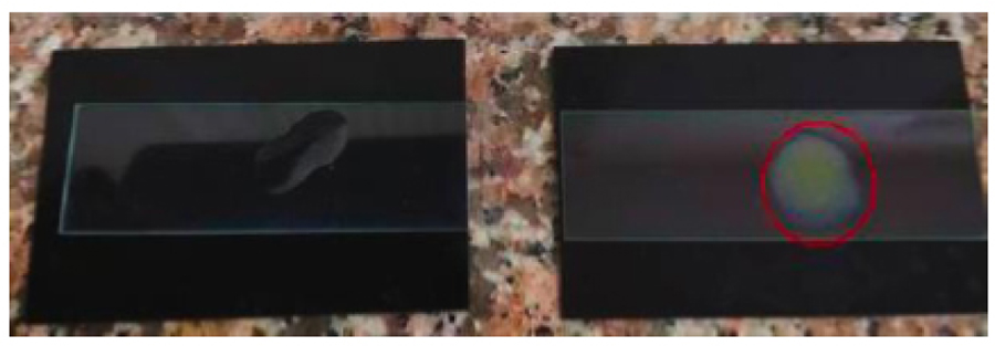
Coagulase test was used for differentiating staphylococcus aureus (+) from Coagulase (-) Staphylococcus (CONS). Coagulase is an enzyme produced by S. aureus that converts (soluble) fibrinogen in plasma to (insoluble) fibrin. The transparent appearance of a coarse clumping (Agglutination) of cocci within 10 seconds suggested the presence of S. aureus (Fig. 10). In contrast, the absence of clumping or any reaction was suggestive of the presence of S. epidermis and S. sapropyticus.
3.3.3. Indole Test Tube Assay
Indole test is a biochemical test performed for the presence of gram (-) bacteria. It measures the ability of the organism to convert tryptophan into indole. In the test, the development of pink to red color on the top alcohol layer shows a positive test (Kovacs positive) reaction. It suggests the presence of Citrobacter koseri, Klebsiella oxytoca, Proteus vulgaris as shown in Fig. (11). In contrast, colorless or light yellow color suggests negative reactions (Kovacs negative). It suggests the presence of Klebsiella pneumonia, Kitrobacter freundii, Proteus mirabilis.
3.3.4. Urease Test
The urea test was performed to identify microbacteria that are capable of hydrolyzing urea using the enzyme Urease. It is commonly used to distinguish the genus Proteus from other enteric bacteria. The appearance of yellow to magenta or bright pink color in 15 min to 24 hours is said to be urea (+ve) (Fig. 12). It suggests the presence of Proteus spp, Helicobacter pylori, Cryptococcus spp, Corynebacterium spp, Yersinia spp, Brucella spp. While the appearance of yellow color (unchanged) is said to be Urease (-ve). It suggests the presence of Escherichia, Shigella, Salmonella.
3.4. DNA Quantification Results
The DNA extraction and quantification showed the positive confirmation of microbial DNA extracted from the culture plates prepared from the containers. Thus, it can be suggested that a higher quantity of Microbial DNA contamination hinders the DNA typing technique by producing its metabolites or by-products inside the samples. These metabolites may also act as inhibitors of the PCR process, interfering with the electrophoresis process during sequencing reaction or leading to undesired target sequence amplification. These microbial contaminated or non-sterilized containers decreased the overall expression of indigenous host cell’s DNA (human DNA). Table 4 shows details of the amount of microbial DNA quantified from the different containers used for evidence collection (recovered from the police station).
Extraction techniques for deficient quantities of DNA that are human-specific are not known or not readily available. Therefore, the presence of microbial DNA in these extracts becomes unavoidable. Studies have shown that various microbial DNAs are sometimes amplified and generate non-specific PCR products that occupy true locus positions resulting in the wrong interpretation of the results. Thus, standard protocols should be followed to minimize the microbial contamination at every step of processing a crime scene, primarily those involving the presence of biological evidence. Due care should be taken in the collection, transport, storage of samples up to the analysis of the samples [23].
| Sample |
Conc. (ng/ul) |
A260(10mm path) | A280 (10mm path) | 260/280 ratio |
|---|---|---|---|---|
| Sample 1 -40ml Screw-capped Vials (White Cap) |
87.9 | 1.758 | 0.979 | 1.7957 |
| Sample 2 -20ml Screw-capped Vials (Blue Cap) |
227.1 | 4.543 | 2.367 | 1.9565 |
| Sample 3 -10ml Screw-capped Vials (White Cap) |
1511.0 | 30.221 | 15.540 | 1.9447 |
| Sample 4 -40ml Screw-capped Vials (Green Cap) |
846.6 | 16.932 | 8.566 | 1.9766 |
| Sample 5 -30ml Screw-capped Vials (Red Cap) |
1928.8 | 38.575 | 20.120 | 1.9172 |
4. DISCUSSION
Maintaining the purity and integrity of the physical evidence (especially biological evidence) found at the crime scene is quite a challenging task. If not done properly and hygienically, it might hamper the scientific conclusions that in succession may mislead information being presented in the court of law. When proper collection methods and sterilized containers are not utilized that should be underlined because it spoils the sensitive information retain in the sample. Nevertheless, there are approved guidelines that suggest using screw cap plastic containers for the collection of biological samples during clinical and forensic analysis [24]. However, these guidelines do not clarify standard operating procedures to avoid the chances of container contamination during the sample collection procedure. Furthermore, the potential for container microbes contaminations (fungi, bacteria, virus) has been a low concern area of law enforcement and forensic practitioners, in general, ever since the evidence was first analyzed. However, the potential impact of evidence contamination upon the outcome of a criminal investigation has become ever more critical because current scientific analysis techniques are much sensitive towards contamination (for example as forensic DNA analysis).
Using plastic containers for biological evidence collection is more common in India, considering it is inexpensive, handy, and more secure, with less likely chances of breakage. Additionally, there is a lack of desire (sterilizing the containers) to follow the standard protocols before the collection of the biological evidence from the crime scene. These lapses pave the way for the containers to get contaminated with microorganism like bacteria, fungi, and viruses [25]. Interestingly, many studies have suggested the association of microbiome with the crime scene and evidences present at the crime scene (skin and biological tissue, clothing’s, computer keywords and other surfaces, gloves, spitting, air). This linkage might be further used for the identification of an individual [15, 26, 27]. With such a surplus presence of microbes, the chances of contamination of biological samples might also be higher. In the present study, we also found exogenous bacterial contaminations inside the containers (Non-sterile). Although, containers were sealed, packed and opened just before the time of evidence collection. Our staining results might suggest the presence of Gram-positive bacteria (Staphylococcus, Bacillus, Corynebacterium, Clostridium) and Gram-negative bacteria (E. coli, Enterobacteriaceae, Pseudomonas, Salmonella, Shigella, Stenotrophomonas, Bdellovibrio, Acetic acid bacteria). We perform biochemical testsin order to identify the specific characteristic of microbacteria inside the culture. These tests suggest the reactivity and nature of bacteria. The Catalase and Coagulase test suggested staphylococcus aureus, Staphylococcus epidermis and Staphylococcus sapropyticus in the microbial culture. The Indole test suggested the presence of Citrobacter koseri, Klebsiella oxytoca, and Proteus vulgaris. Some of the urea metabolizing bacteria (Proteus spp, Helicobacter pylori, Cryptococcus spp, Corynebacterium spp) might be present in the microbial culture.
This study becomes quite significant in the Indian context because of the prevailing environmental conditions (Temperature, humidity, air quality, pH) in a tropical region that favours for airborne and soil inhabitant bacterial growth during the spring sessions. Due to high moisture contents, degradation rate, and high bacterial growth in biological evidence, it is difficult to retrieve and identify genetic material (RNA, DNA) from the trace sample [3, 4]. While DNA isolation is an essential and the first step in molecular, biological applications and its efficacy depends mainly on the source, type, and purity of the sample. Different technical caveats challenge and distort the DNA extraction process; some of the main are improper handling, preservation, inhibitors, complex microbial matrix, sludge, and soil contamination [28]. Since the biological sample of forensic importance is highly rich in moisture and nutrient contents. These nutrients and moisture conditions aid to growth and survive both endogenous and exogenous microorganism. The dead host cells (less immunised) doesn’t inhibit to growth of the microbes that result into rapid microbial degradation and more by-products formation through physiological process. The microbes and their by-products may hinder DNA profiling and will give biased results either by degrading or modifying the DNA, inhibiting PCR reaction or by interfering with sequencing reaction. As a result, necessary measures are needed to minimize microbial contamination at every step of DNA profiling, starting from sample collection to transport storage and downstream processing [23]. Thus packaging equipment or means must also be free of contamination. Furthermore, it has been reported that due to the high burden of crime rate, untrained police investigators collect the biological samples without taking proper care.
CONCLUSION
This study recommends that there should be proper maintenance of the chain of custody to collect and preserve the evidence properly to prevent degradation or contamination in the biological evidence. The biological evidence needs extra care during collection and packing. At crime scene, when containers are left open, just before the collection, the chance for contamination increases dramatically. Improper container handling might also increase the contamination inside the containers. So, regular training workshops for investigators and forensic specialists must be conducted to ensure proper handling of biological evidence, from the scene to storage that ultimately reduces the risk for contaminations and addresses correct issues of an investigation. Freshly manufactured, screw-capped, sterilized containers must be used to pack evidence, particularly in biological evidence. The packaging equipment must also be free of contaminations. These can be easily accomplished by keeping all the packaging supplies in one case and handling them carefully. However, the study is required to profile the microbial diversity and their functional attributes which are responsible for the degradation of biological samples or significant contaminations. Consequently, metagenomics will be an appropriate way to identify the whole array of microorganisms and their role in the degradation of samples of biological origin.
ETHICS APPROVAL AND CONSENT TO PARTICIPATE
Not applicable.
HUMAN AND ANIMAL RIGHTS
No animals/humans were used for studies that are the basis of this research.
CONSENT FOR PUBLICATION
Not applicable.
AVAILABILITY OF DATA AND MATERIALS
The data supporting findings of this study is present within the article.
FUNDING
None.
CONFLICT OF INTEREST
The authors declare no conflict of interest, financial or otherwise.
ACKNOWLEDGEMENTS
All authors would like to say thank to Sector 58, Noida Police Station for providing the container samples, and Shriram Singh Multispecialty Hospital, Delhi. Also, acknowledge to Thermofisher, Noida (UP) for providing the Training and Laboratory facilities for sample analysis. All authors are thankful to CTM-IRTE, Faridabad, India, for providing the opportunity to work and their support and interest

