All published articles of this journal are available on ScienceDirect.

Evaluation of Biofilm Formation and Anti-biofilm Properties of Peganum Harmala and Crocus Sativus in Shigella Flexneri Clinical Isolates
Authors Info & Affiliations
Abstract
Background:
Biofilm formation causes many serious problems in the treatment of bacterial infections. In addition, chronic infections due to biofilm formation can pose a huge burden to the health care systems. Also, many bacteria are biofilm producers as an important strategy for pathogenicity. Furthermore, the traditional use of herbal medicines such as Peganum harmala and Crocus sativus in Iran is interesting.
Objective:
The purpose of the current study was to investigate the biofilm formation in Shigella flexneri clinical isolates and to evaluate the anti-biofilm properties of P. harmala and C. sativus on Shigella flexneri clinical isolates.
Methods:
For the study purpose, Thirty S.flexneri clinical isolates were collected from Ahvaz, Iran. Then, the collected bacteria were subjected to biofilm formation assay. Afterward, P. harmala and C. sativus were applied as an anti-biofilm formation in S. flexneri.
Results & Conclusion:
Our results demonstrated that a significant number of samples were identified as strong biofilm producers. Then, P. harmala and C . sativus in a concentration of 30μg/ml and 60μg/ml were able to eradicate a strong biofilm formation in S. flexneri, respectively. In addition, it seems that more extensive studies and in vivo research should be done to confirm their properties.
1. INTRODUCTION
Despite, there are many virulence factors in bacteria; the role of biofilm is notable [1]. The biofilm structure has many complications on human health and environment [2].In addition , numerous chronic infectious diseases are caused by biofilm formation in bacteria [3]. Many bacteria are biofilm producers, among them some true pathogens are more considerable for their biofilm formation [4,5].
Though, biofilm formation was investigated in different species of bacteria, including Pseudomonas aeruginosa, Enterobacteriaceae etc [6], Shigella species is considered in some countries to have the ability to produce biofilm [7]. However, S. flexneri may also be one of the biofilm producers other than Shigella species. Many studies have investigated the intracellular survival and virulence factor in S. flexneri; but there is a big gap in the pathogenesis and intracellular survival of this bacterium [8].
Certainly, biofilm formation is a mechanism in bacteria, which can promote bacterial resistance [9]. Furthermore, the use of antibiotic treatments against the biofilm structure in bacteria is one of the main challenges in medical science [10]. New drug discovery can be a good choice for the eradication of biofilm formation. Also, many studies have shown that different types of medicinal plants can be considered as an effective weapon against infectious diseases [11]. In the meantime, P. harmala is originally a native Asian plant. This plant belongs to the Nitrariaceae family. P. harmala is traditionally used to treat many infectious diseases [12]. Besides that, Crocus sativus is a native plant of Iran and used in traditional medicine in this country. The essential oil of this plant has antibacterial effects [13]. Due to these reasons, in this study, biofilm formation by S. flexneri isolates was investigated and the anti-biofilm properties of P.harmala and C.sativus on S. flexenarii clinical isolates were evaluated.
2. METHODS
2.1. Bacterial Collection and Identification
A total of thirty S. flexneri clinical isolates were prepared at the Microbiology Research Center, Ilam University of Medical Sciences. Ilam, Iran. Then, S. flexneri were obtained from Ahvaz, Iran, by a a standard method [14,15].
2.2. Cell Culture
The P. harmala and C. sativus ethanolic extracts were applied to determine their cytotoxic effect on a vero cell line. Then, the MTT assay was performed by the MTT assay kit (Sigma, United States).
2.3. Toxicity Assay
The cells were inoculated in 96-well microplates and cellular density was determined. Then, the cells encountered different concentrations of the P. harmala and C. sativus extracts. The MTT assay was performed and the absorbance of the transformed dye was measured at a 600nm wavelength.
2.4. Biofilm Formation Assay
Initially, 0.5 McFarland solution of S. flexneri was prepared. Then, we inoculated 200 uL of broth media (LB broth) with a 0.5 McFarland solution of S. flexneri in 96 microplates for the evaluation of biofilm formation. Henceforth, the culture incubated for 24 hours at 35°C, so, the experiment was performed in triplicate. LB broth without S. flexneri was a negative control.
2.5. Semi-quantification of Biofilm Biomass
In this study, we used the methodology defined by Mowat et al. [16].
2.6. Analysis of Biofilm Formation
The ability of biofilm formation in all S. flexneri isolates was measured by absorbance in the crystal violet stain. In addition, the capacity of all of the strains to form a biofilm was compared with biofilm-forming S. flexneri controls. Further- more, we measured biofilm formation for each sample by analyzing the absorbance of the crystal violet. In this process, each isolate can create a biofilm mass in 24 hours which is eventually compared with the control. Finally, the isolates were divided into three categories based on biofilm formation. These groups included biofilms with 75% of the biomass of the positive control, moderately adherent biofilms with 25-75% biomass or weak biofilms with 25% of the biomass of the positive control.
3. RESULTS
3.1. Biofilm Formation by S. flexneri
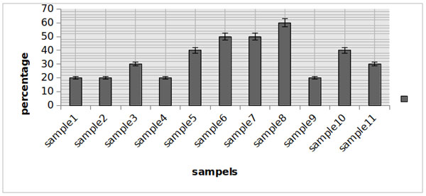
Initially, the bacteria were confirmed by phenotypic methods. Furthermore, we discovered biofilm formations as a significant factor in S. flexneri clinical isolates; while the largest number of clinical isolates with a strong biofilm structure (n=11). In some S. flexneri clinical isolates, a moderate biofilm formation was also significant (n=10). Nevertheless, S. flexneri isolate was also observed with a weak biofilm formation (n=8). In addition, strains with no biofilm were very low and negligible (n=1). These results are summarized in Fig (1).

3.2. P. harmala as An Anti-biofilm Formation in S. flexneri
The IC50 of P. harmala was 35 μg/ml. In this study, eleven isolates (36.66 percentages of samples) were observed to be able to produce a strong biofilm. Different concentrations of P. harmala was tested for all of them. P. harmala in a concen- tration of 30μg/ml could eradicate the biofilm formation (Fig. 2).

3.3. C. sativus as An Anti-biofilm Formation in S. flexneri
The IC50 of C. sativus was 100 μg/ml. Different concen- trations of C. sativus were tested for eleven isolates (36.66 percentages of samples) with a strong biofilm formation. C. sativus in a concentration of 60μg/ml easily eradicated the biofilm of S. flexenary (Fig. 3).
4. DISCUSSION
The biofilm formation causes many serious problems in the development of effective therapies for the treatment of infectious diseases [17]. However, the inherent ability of biofilm production in some bacteria has created many challenges in medical science [18]. Also, biofilm formation can cause widespread complications in the treatment of diseases and in the maintainence of human health [19].
In addition, biofilm formation is a community of microorganisms, which results in many infections and diseases causing problems at biological and environmental level [20].
CONCLUSION
In fact, one of the main mechanisms of bacterial survival in different environments is the ability to produce biofilms [21]. Moreover, bacteria that have the capacity to create biofilms can escape the host immune system, and therefore, cause chronic infections [22]. Meanwhile, S. flexneri employs several strategies to escape the immune system; one of the most important strategies is the ability to produce a biofilm [23]. In some studies, the effective factor in biofilm formation by S. flexneri was investigated [24]. Also, in several studies, S. flexneri infection was investigated but there remained a huge and significant gap in our knowledge for how to make S. flexneri capable of surviving in stress conditions [25]. Our data demonstrated that biofilm formation is a significant factor in S. flexneri clinical isolates. Furthermore, our results declare that medicinal plants can be used as a suitable candidate for the treatment of biofilm formation caused by S. flexneri. However, it seems that in vivo studies and more extensive studies in the field are necessary.
ETHICAL APPROVAL AND CONSENT TO PARTICIPATE
The current research approved by Ethical committee of Ilam University of Medical Sciences, Iran.
HUMAN AND ANIMAL RIGHTS
Not applicable.
CONSENT FOR PUBLICATION
Not applicable.
AVAILABILITY OF DATA AND MATERIALS
Not applicable.
FUNDING
None.
CONFLICT OF INTEREST
The authors declare no conflict of interest, financial or otherwise.
ACKNOWLEDGEMENTS
We would like to thank the clinical microbiology research center, Ilam University of medical sciences for the strains used in this study.

