All published articles of this journal are available on ScienceDirect.

Non Tuberculosis Mycobacterium, E. coli and E. faecalis from Biofilm in Drinking Water Distribution Systems from Selected Sites of Addis Ababa
Authors Info & Affiliations
Abstract
Background:
The decline in microbial quality of drinking water may be attributed to many factors among which the presence of biofilm within the distribution system is the major cause of contamination. Drinking water distribution systems provide an oligotrophic environment, for post-treatment recovery and regrowth of microorganisms including the opportunistic Nontuberculosis Mycobacterium (NTM).
Objective:
The aim was to look for opportunistic non tuberculosis mycobacterium and indicator organisms of fecal contamination from biofilm in drinking water distribution pipeline from selected sites of Addis Ababa.
Materials and Methods:
A total of 40 biofilm samples were collected from two sub-cities of Addis Ababa. Biofilm samples were taken from the inner surfaces of the get valve and water meter. For the detection of E. coli and E. faecalis, diluted biofilm samples were filtered, then it was incubated on respective culture media. For non-tuberculosis mycobacterium, the homogenized biofilm sediment was processed using the standard SD bio line method, whereby, The processed sediment was inoculated to appropriate solid and liquid culture media. The DNA extraction was conducted by chemical lysis followed by PCR amplification, from the grown colonies on LJ media (Löwenstein–Jensen). The identification of Mycobacterium species was performed by reverse hybridization using a membrane strip and an enzymatic color reaction.
Results:
From the total biofilm samples, 14 out of 40 (35%) were positive for mycobacteria species. M. gordonea was the most prevalent specie of Mycobacterium, whereby 8/14 (57.1%) of the isolates were from this species followed by M. fortuitum 1/14 (7.14%). About (35.7%) 5/14 of the genus Mycobaterium were unidentified species. Indicator organisms of fecal contamination (E. coli and E. faecalis) were found in 3/40(7.5%) and 6/40(15%) respectively. There was no statistically significant association between nontuberculosis mycobacterium and the indicator organisms at p value of 0.01.
Conclusion:
The study has highlighted that the occurrence of NTM in drinking water distribution in a significant proportion. M. gordonae was found to be the most dominant species of nontuberculosis mycobacterium found in the distribution line biofilm samples.
1. BACKGROUND AND JUSTIFICATION
Public drinking water distribution system in the urban setting of developing countries supports millions of people in providing “safe water”. There are different factors which increase the risk of microbial contamination of piped treated water such as stagnation of water and low chlorine residuals [1]. Drinking water distribution systems provide an oligotrophic environment for the survival, post-treatment recovery and regrowth of bacteria [2]. Such kind of environment assists in the formation of biofilm which will shelter and protect microorganisms from disinfection [3].
The genus mycobacterium comprises both the strict pathogens such as M. tuberculosis and M. leprae while for Nontuberclosis Mycobacteria (NTM), the Mycobacterium Avium Complex (MAC) which are the major opportunistic pathogens. NTM is considered as an important group comprising of opportunistic and strict pathogens such as M. avium complex [4]. There are more than 150 NTM species currently recognized, about 25 of them have been associated with NTM diseases in humans and/or animals [5]. The most frequently isolated species of NTM are Mycobacterium gordonae, Mycobacterium kansasii and Mycobacterium chelonae from clinical cases [6, 7].
The ability to tolerate different situations such as variation in temperature, pH, and the ability to resist to disinfectants makes NTM survive better than the rest of bacteria living in drinking water [5]. MAC has a high resistance to chlorine; it can tolerate chlorine concentrations of 0.05-0.2 mg/L found in tap water [8, 9], among which M.abscssus, M. gilvum. M.gordonae and M. magreitance have been associated with municipal water supplies.
The aim of this study was to look for opportunistic NTM and other indicator organisms (E.coli, E.faecalis) of fecal contamination and their association with biofilm in distribution pipeline from selected sites of Addis Ababa.
2. MATERIALS AND METHODS
2.1. Biofilm Sample Collections
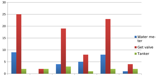
A total of 40 biofilm samples were collected from two sub-cities of Addis Ababa. The biofilm sample was taken from the inner surfaces of the faucets of the get valve and strainer/filter of water meter depending on the suitability for taking a sample using a sterile cotton-tipped swab moistened in 1ml sterile normal saline solutions. The area of biofilm removed in this way was approximately 1cm2. Collected samples were transported to the laboratory in an ice box and analyzed within 12 to 18 hr. From the total biofilm samples, 10 were taken from the strainer/water filter of the water meter, 27 from the get valve and 3 from the tanker or reservoir.
2.1.1. Laboratory Analysis of Biofilm Samples
Biofilm samples were dispersed by vigorous vortexing in sterile physiological saline water. The homogenized samples were analyzed to determine the detection of E. coli, E. faecalis and mycobacterium spp.
2.1.2. Detection of E. coli
The homogenized swab samples were diluted in peptone water filtered using cellulose membrane 0.45µm. The filtered membrane was transferred onto Tryptone Soya Agar (BBL) plates using sterile forceps and incubated for 4 h at 37°C. After 4 hrs of incubation, the membrane was transferred to Tryptic Bile Agar (BBL) at 44°C for 16-24 hr incubation. Finally, an indole test was done with the addition of 1-2 drops of James reagent on the colonies grown on the membrane.
2.1.3. Detection of E. faecalis
Filtered membrane was transferred onto, Slantz Bartley agar (Oxoid) and incubated at 37ºC for 16-24 hours; the formation of brown- red colonies was regarded as presumptive positive. Typical black colonies formation on Bile Esculin Agar plates (BBL) after 4-12hr incubation at 44 ºC was regarded as E. faecalis [10].
2.1.4. Culture and Identification of NTM
The homogenized biofilm sample was processed using N-acetyl-L-cysteine-sodium hydroxide (4%NaOH-NALC) method using the standard method of (SD bioline). The processed sediment was inoculated to appropriate liquid and solid culture media, 7ml Middlebrook 7H9 Mycobacterium Growth Indicator Tube (MGIT) liquid culture media incubated to Bactech MGIT 960 (Becton Dickinson Microbiology Systems, Cockeysville, MD, USA) until it flags positive or is confirmed by the characteristic growth and Lowenstein-Jensen (LJ) Solid Culture media. All culture-positive workup was done to identify using MPT64 Ag test [SD bioline] serological test, true positivity, whereby all true culture positive isolates were further processed for molecular typing.
2.1.5. Genotyping of NTM
Colonies of mycobacterium grown on LJ were prepared by suspending a loopful of the bacteria in 1 ml distilled water before extraction. For DNA extraction, the GenoLyse® kit and 500 micro liters of liquid culture was used. DNA extraction was performed by chemical lysis followed by heating to 95 °C for 5 min, centrifuged at 13400 g for 5 min and the supernatant was used for the assay. All the reagents needed for amplification were included in the Amplification Mixes A and B. After thawing, it was spin down AM-A and AM-B briefly and was mixed carefully by pipetting up and down in a room free from contaminating DNA. The DNA solution was added in a separate working area. Hybridization was performed by manually washing in an automated shaking water bath which was prewarmed at 45°C. The solutions HYB and STR were prewarmed to 37-45°C before use and were mixed. Using a suitable tube, the solutions were diluted to Conjugate Concentrate (CON-C, orange) and Substrate Concentrate (SUB-C, yellow) 1:100 with the respective buffer, It was mixed well and bring to room temperature. For each strip, 10 µl concentrate was added to 1 ml of the respective buffer to dilute CON-C before each use. After a final washing step, strips were air-dried and fixed on a paper; only bands whose intensities were as strong as or stronger than the universal control line were considered Mycobacterium CM VER 2.0 [10].
3. RESULTS
A large number of biofilm samples were collected from household 25/40(62.5%), and community tap water 5/40 (12.5%) presented in Fig. (1).

| Sample Source |
Isolates (percentage) |
Mycobacteruim spp. |
|---|---|---|
| Reservoir | 1(7.14%) | Mycobacterium spp |
| Household | 3(21.4%) | Mycobacterium spp |
| 4(28.5%) | M.gordonae | |
| 1(7.14%) | M.fortunae | |
| Community water | 1(7.14%) | M.gordonae |
| Health Facility | 1(7.14%) | M.gordonae |
| 1(7.14%) | Mycobacterium spp | |
| Community Recreation | 2(14.3%) | M.gordonae |
| Source of sample | Sample Taken From | Isolates (percentage) |
|---|---|---|
| Community water | Get valve | 1(33.3%) |
| Reservoir | Taken from tanker | 2(66.6%) |
3.1. Non-tuberculosis Mycobacterium
In general, out of the total 40 biofilm samples collected, positive isolates of mycobacterium species were found in 14(35%). There was a significant variation between the two sub-cities in terms of the occurrence of NTM which was 2/14 (14.3%) and 12/14 (85.0%) in the other sub-city. The majority of Mycobacterium 8/40 (20%) species were isolated from a household.
Among the isolates of NTM, M.gordonae was the most abundant species isolated 6/14(43%). There was only one species of M. fortuitum which was isolated from the household, the rest 3 mycobacterium were unidentified species.
3.2. E. coli
E. coli was isolated in only 3/40(7.5%) of the biofilm samples, from which, 2/3(66.6%) of the isolates were found from tanker/reservoir, while 33.3% (1/3) from community water (Table 2).
3.3. E. faecalis
Out of the total biofilm samples collected, 6/40(15%) were positive for E. faecalis. The distribution of the isolated E. faecalis by the source is shown in Table 3.
Pearson correlation coefficient showed that the correlation between NTM and indicator organisms was not statistically significant at 0.01. While for E. coli and E. faecalis, there was statistically significant association at 0.01 levels (Table 4).
Detection of the three organisms i.e. mycobacterium species with the other two indicator organisms (E. coli and E.faecalis) was found in 1/40 (2.5%) of the biofilm samples. While the co-occurrence of mycobacterium with either of the indicator organisms was 3/40(8%). The probability of finding one of the three organisms from biofilm was 15/40 (37%) (Fig. 2.
4. DISCUSSION
A significant percent of pathogen exists in a viable but noncultivable state, unable to grow on artificial growth media but alive and capable of renewed activity, and therefore being hygienically relevant [12]. Different literatures suggest that [11] in drinking water systems, the high majority of bacteria, estimated to be 95%, are located attached to the surfaces of pipelines, in the form of biofilm. While only 5% are found in the water phase (sessile phase) and detected by sampling as commonly used for quality control.
Biofilm samples taken from tankers or reservoir were highly predicted to have more pathogens as compared to samples taken from the get valve and water meter. It can be assumed as water is kept for a period of time, there is always an environment for the formation of biofilm, its survival and multiplication of microorganisms.
In a similar study conducted in the USA Mycobacteria were isolated in 16 (38%) of 42 public drinking water distribution systems [13], while in Finnish study, they found out that the isolation frequency of mycobacteria from drinking water distribution system samples was 35% and increased up to 80% at the most distal sites of the waterworks [14]. In this study Mycobacterium was isolated in 35% of the biofilm samples.
The species M. avium, M. chelonae, M. fortuitum, M. gordonae, M. kansasii, and M. xenopi are the most frequently reported mycobacteria occurring in drinking water [15]. In this study, M. gordonea, (creamy yellowish color) was the most prevalent species, while M. fortuitum (a smooth grayish color) was the second most prevalent species of NTM.
Pearson correlation coefficient at p-value of 0.01 showed that there was no statistically significant correlation between NTM and indicator organisms of fecal contamination. But there was is significant correlation between E. coli and E. faecalis at p-value of 0.01 with the Pearson correlation coefficient of 0.504.
In a similar study but from drinking water it was found that M. gordonea was one of the dominant species isolated among the other species of NTM [15].
| Source of sample | Sample taken from | Isolates (Percentage) |
|---|---|---|
| Household | Getvalve | 2 (33.3%) |
| Reservoir | Tanker | 2(33.3%) |
| Community Recreation |
Filter of water meter | 1(16.6%) |
| Community tap water |
Filter of water meter | 1(16.6%) |
| NTM | E. coli | E. faecalis | ||
|---|---|---|---|---|
| NTM | Pearson Correlation |
1 | -.070 | .214 |
| Sig. (2-tailed) | - | .668 | .185 | |
| N | 40 | 40 | 40 | |
| E.coli | Pearson Correlation |
-.070 | 1 | .504** |
| Sig. (2-tailed) | .668 | - | .001 | |
| N | 40 | 40 | 40 | |
| E.faecalis | Pearson Correlation |
.214 | .504** | 1 |
| Sig. (2-tailed) | .185 | .001 | ||
| N | 40 | 40 | 40 | |
CONCLUSION
This is of the first kind of study conducted in drinking water in Ethiopia to look for the diversity of NTM and indicator organisms drinking water distribution from biofilm. The study has highlighted that biofilm is one of the potential sources of contamination in the drinking water distribution system. Further research work on a large scale on the health impact and significance of the environmental non Mycobacterium (NTM) should be conducted .
ETHICS APPROVAL AND CONSENT TO PARTICIPATE
The protocol and methods employed were reviewed and approved by the Institutional Review Board, Faculty of medicine, Mansoura University, Mansoura, Egypt.
HUMAN AND ANIMAL RIGHTS
No animals/humans were used for studies that are the basis of this research.
CONSENT FOR PUBLICATION
Informed consent was obtained from all the participants.
AVAILABILITY OF DATA AND MATERIALS
Not applicable.
FUNDING
None.
CONFLICT OF INTEREST
The authors declare no conflict of interest, financial or otherwise.
ACKNOWLEDGEMENTS
Declared none.

