All published articles of this journal are available on ScienceDirect.

Isolation of L-Asparaginase-Producing Actinomycetes from Soil in Qom: Characterization and Anticancer Activity of L-Asparaginase Against MCF-7 Breast Cancer Cells
 Abbas Morovvati1
Authors Info & Affiliations
Abstract
Introduction
Actinomycetes play an important role in the pharmaceutical and biotechnology industries due to their ability to produce secondary metabolites. Among the most important enzymes they produce is L-asparaginase, a key drug used to treat blood cancer such as acute lymphoblastic leukemia. However, its production is costly and resource-intensive, highlighting the need for the identification of new sources and optimization of its production. The present study aimed to isolate and identify L-asparaginase-producing actinomycetes collected from soil samples in Qom City, Qom, Iran, optimize the conditions for enzyme production, and assess its anticancer effects on the MCF-7 Breast Cancer (BC) cell line in vitro.
Materials and Methods
In this laboratory study, soil samples were collected from various locations in Qom. Specific culture methods were used to isolate L-asparaginase-producing actinomycetes. The production conditions of the enzyme were optimized with respect to temperature, pH, and carbon and nitrogen sources. The effect of L-asparaginase on the MCF-7 cell line was tested for anticancer activity by the MTT method.
Results
Among the 65 isolated colonies, three colonies showed the ability to produce L-asparaginase, and the superior strain with higher activity, Streptomyces flavoviridis, was identified. The specific activity of the enzyme increased to 415.25 mg protein/unit after purification, and the analysis by SDS-PAGE indicated a 30 kDa protein band. The enzyme extracted inhibited the growth of MCF-7 BC cells in a dose- and time-dependent manner, with IC50 of 25.46 and 16.54 µg/mL at 24 and 48 h, respectively.
Discussion
The findings showed that actinomycetes are suitable sources for producing L-asparaginase with anticancer effects.
Conclusion
The strains identified in this study could be a suitable solution for the industrial production of this enzyme with lower cost and greater effectiveness.
1. INTRODUCTION
The large family of actinomycetes comprises various species of bacteria that are widely found in nature, particularly in soil. The diversity and abundance of actinomycetes in different types of soils are influenced by geographical regions, agricultural practices, and soil minerals [1, 2]. Despite the high presence of actinomycetes in various soils, only a small number are cultivated during isolation processes [3]. Currently, soil is the most promising source for isolating actinomycetes; however, relatively few studies have been conducted in this area in our country [4].
Many efforts have been made to produce antibiotics from bacteria in the family Actinomycetaceae, especially those belonging to the genus Streptomyces. However, less attention has been paid to other bacteria in this family due to the challenges associated with their isolation and susceptibility to growth inhibition by other environmental bacteria [5, 6]. On the other hand, due to the increasing clinical emergency needs for new treatments and the rising issues related to chemotherapy, such as the emergence of antibiotic-resistant bacteria and the growing number of patients with weakened immune systems, as well as the diversity of non-infectious diseases such as cancer, there is a pressing need to isolate new antibiotics and bacterial metabolites with antitumor properties. Additionally, enzymes with anticancer properties, such as asparaginase, are being sought [7, 8].
Several bacteria capable of producing asparaginase have been identified, but their low production levels have led to ongoing activities to find microorganisms that can produce higher quantities of this enzyme. If enough of these enzymes can be obtained, valuable research can be conducted to treat various types of cancer. In Iran, however, limited research has been conducted on the isolation and purification of asparaginase from actinomycetes [9, 10]. The aim of the present study was to isolate and identify L-asparaginase-producing actinomycetes collected from soil samples in Qom Province (Qom, Iran), to optimize the conditions for enzyme production and evaluate its anticancer activity against the MCF-7 BC cell line in vitro.
2. MATERIALS AND METHODS
2.1. Sample Collection
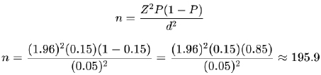
Soil samples were collected from the Qom Salt Lake area because saline and environmentally stressful habitats may contain physiologically adapted actinomycetes with diverse metabolic capabilities and the potential to produce novel bioactive enzymes, including L-asparaginase. According to the results obtained from research, the prevalence rate of actinomycetes in these environmental soil samples was found to be 27%. To estimate this rate from the detected samples at a 95% confidence level and a 5% error rate, we selected 284 soil samples based on the sample size formula [11].
After determining the pH of the collected soil sample, 1 g of the soil was dissolved in 10 mL of distilled water. Initial cultivation was performed on bottom agar and chitin agar media. After 14 to 21 days of incubation at 25 to 28 °C, a microscopic examination was performed for evaluation. In the next step, each type of colony was cultured on glucose- yeast extract agar plates. After incubation [12], suspected bacteria were isolated from soil samples for the initial identification of the bacterial genus and species using phenotypic tests.
2.2. Enzyme Activity Assay
Enzyme activities were evaluated using standard biochemical tests, including growth ability in lysozyme broth, casein, tyrosine, xanthine, hypoxanthine, and gelatin hydrolysis tests, as well as urease, nitrate reductase, and L-asparaginase (asparagine amidohydrolase) activities.
2.3. Extraction and Enzyme Production
Primary enzymes were extracted by the solid-state fermentation method [13]. To do this process, we added soybean meal (5 g) to 0.1 M sodium phosphate buffer (10 mL) in a 250 mL Erlenmeyer flask and autoclaved at 121°C for 15 min. After cooling the medium, 3 mL of the bacterial suspension prepared previously was added to the medium. This mixture was left to incubate at 37 °C for 4 days without shaking. Sodium phosphate buffer (90 mL) was then added to the enzyme, and it was precipitated by centrifugation at 8000 rpm for 20 min. An alternative method was the inoculation of 1 mL of the spore suspension into the Erlenmeyer flasks containing 50 mL of preculture medium. This mixture was shaken (220 rpm) at 28 °C for 48 h. The preculture was subsequently transferred to the fermentation medium to obtain 5% (v/v) of it and cultured at 27 °C for 8 days on a shaker at 220 rpm. Thereafter, the pH of the flask was measured after fermentation, and the supernatant was separated by centrifugation at 4000 rpm for 20 min. Extraction was performed with ethyl acetate (2:1, v/v), and the solvent was evaporated at 39 °C using a rotary evaporator.
2.4. Enzyme Purification
The activity of the enzyme L-asparaginase was determined by a colorimetric method. For this purpose, 0.04 M of L-asparagine, crude enzyme extract, and 0.5 M of Tris-HCl buffer (0.5 mL each) were mixed and then placed in a water bath at 37 °C for 15 min. Next, 0.5 mL of 1.5 M trichloroacetic acid solution was added to the mixture, followed by a 30 min centrifugation at 10,000 rpm for 30 min. Afterwards, 0.1 mL of Romand's liquid was added to Nessler's reagent (0.2 mL) and distilled water (3.7 mL). The mixture was then allowed to stand at room temperature for 15 min. Ammonia released was quantified by measuring with a spectrophotometer at 480 nm. Activity of the enzyme was expressed in international units and calculated by the ammonium sulfate standard curve [14]. Enzyme purification can be performed by various methods, such as dialysis, ammonium sulfate precipitation, Sephadex G100, GM cellulose, DEAE Sephadex chromatography, and CM-Sephadex C50 ion-exchange chromatography. Purification was carried out at 0 to 4°C for ammonium sulfate purification. Ammonium sulfate powder was added to the liquid culture until it reached 40% saturation and incubated at 4 °C overnight. After that, the saturation was increased to 80% by adding ammonium sulfate. The next step was centrifugation at 8000 rpm for 10 min, and the obtained precipitate was dissolved in Tris-HCl buffer, followed by dialysis in Tris buffer for 8 h. In this study, the final purification of the enzyme was performed using an HPLC system with Sephadex G-200, using ammonium sulfate and Tris-HCl buffer. The molecular weight of the enzyme was also measured by SDS-PAGE.
2.5. Optimal Conditions for Enzyme Activity
To investigate and determine the optimal conditions affecting enzyme production and activity, including temperature, pH, and carbon source, various experiments were designed and performed [15]. After culturing BHI agar at 33 °C for 24 h, strain S1 was prepared as a 0.5 McFarland suspension, and 2 mL of it was inoculated into the enzyme production liquid medium. To determine the optimal temperature, samples were incubated in a shaking incubator at a speed of 151 rpm at different temperatures (30, 35, 40, and 45 °C) for 44 h. Then, the enzyme activity was measured. To study the effect of pH, the pH of the medium was adjusted to 6, 7, 8, and 9 before sterilization. After inoculation, the flasks were incubated at 151 rpm at 30 °C for 44 h, and the enzyme activity was determined. To evaluate the effect of carbon sources, the enzyme production medium was supplemented separately with glucose, xylose, maltose, and lactose. After inoculating strain S1 and incubating the samples under the same conditions, the enzyme activity was measured quantitatively.
2.6. Sodium Dodecyl Sulfate-Polyacrylamide Gel Electrophoresis (SDS-PAGE)
SDS-PAGE electrophoresis was conducted to determine the apparent molecular weight and to test the purity of the enzyme in a vertical electrophoresis system with 7.5% separating gel and 3.5% consolidating gel [16]. Protein samples were mixed with loading buffer (SDS, mercaptoethanol, Tris-HCl [pH 6.8], glycerol, and bromophenol blue) and heated at 100 °C for 10 min. The samples were then placed into the gel and subjected to electrophoresis at 160 V for 4 h. FastGene® BlueEasy Protein Ladder (BlueEasy Prestained Protein Marker, Three-Color, 6.5–270 kDa; [Nippon Genetics Co., Ltd., Japan]) was used as a standard marker to determine molecular weight. The stained gel with Coomassie Brilliant Blue R-250 was destained after electrophoresis. The apparent molecular weight of the protein was determined by comparing the position of the band with that of the markers.
2.7. Measurement of Asparaginase Activity and Production Optimization
A colorimetric method was used to measure the activity of asparaginase [17]. In this technique, a water bath was utilized to incubate the crude extract of the enzyme, Tris-HCl buffer, and asparagine at 37 °C. The reaction was stopped after 15 min when trichloroacetic acid was added. After centrifugation, the supernatant, Nessler's reagent, and distilled water were mixed and preserved at room temperature for 15 min. Ammonia release was determined by spectrophotometer measurement at 450 nm, and enzyme activity was quantified by using the ammonium sulfate standard curve. Growth temperature, pH, carbon sources (glucose, fructose, arabinose, and galactose), nitrogen sources (asparagine, glutamine, peptone, tryptone, and malt extract), and metal ions (Zn2+, Cu2+, and Fe2+) were studied for their effects on asparaginase production over a 72-h period in the ISP5, with modifications to produce the enzyme. The maximum level of asparaginase production was calculated under the optimized conditions, and the optimal growth conditions were recorded.
2.8. Evaluation of the Anticancer Effects of the Enzyme by MTT (Diphenyltetrazolium Bromide) Assay
Cytotoxic effects of the extracted enzyme on MCF-7 cancer cells (Prepared by the Pasteur Institute of Iran) were assessed using the MTT assay [18]. The cells were cultured in DMEM medium and transferred to a 96-well microplate at a density of 3.5 × 10⁵ cells/mL. The extracted enzyme samples were added to the wells after appropriate dilution, and the wells were considered control wells without enzyme. Based on the change in cell shape (granulation, rounding, and wrinkling), the cellular effects were examined under a microscope within 24 to 72 h. MTT was used as a substrate to determine the IC50. After incubating at 37 °C for 4 h, the living cells converted MTT into formazan crystals. To dissolve the formazan, the wells were washed with 110 μL of DMSO or isopropanol, resulting in a purple color. Absorbance was read at 570 nm using a microplate reader, and the IC50 was calculated based on the standard curve.
2.9. Molecular and Phylogenetic Evaluation
DNA of bacterial isolates was extracted using isopropanol precipitation and the cetyltrimethylammonium bromide method [19]. For molecular identification, the 16S rRNA gene was amplified by PCR using specific primers (Table 1). After purification, PCR products were subjected to sequencing. The resulting sequences were examined with Finch TV software, and a consensus sequence was extracted. Phylogenetic analysis was performed through alignment with NCBI reference sequences using MEGA 7 software, which included identification of variable and conserved regions, singleton and phylogenetically informative sites, nucleotide differences, amino acid changes, phylogenetic tree drawing, and sequence type determination.
| Gene | Primer | Sequence | Tm | Product Length |
PCR Cycling Conditions |
|---|---|---|---|---|---|
| 16S rRNA | 27F | 5'AGAGTTTGATCCTGGCTCAG3' | 56.92 | 1480 bp | 95 °C, 4 min; 94 °C, 45 s; 54 °C, 45 s; 72 °C (30 cycle); 72 °C, 10 min |
| 1492R | 5'- GGTTACCTTGTTACGACTT -3' | 52.20 |
3. RESULTS
3.1. Isolation, Screening, and Initial and Molecular Identification of L-asparaginase-Producing Bacteria
After initial linear culture of the samples and examination of plates obtained from different dilutions, a total of 65 isolated and single-growth colonies were identified. These colonies were examined in M9-specific medium for the initial screening of L-asparaginase-producing bacteria. Among the screened colonies, three were selected due to their ability to change the color of the culture medium to pink, indicating L-asparaginase activity. These colonies were further analyzed to measure enzyme activity and to identify the strains. The initial identification of the selected strains was performed using Gram staining and a series of biochemical tests. The results of these tests are presented in Table 2 and included oxidase and catalase tests, growth range at different NaCl concentrations (0–5%), growth pH range (7–10), hydrolysis tests (gelatin, starch, Tween 80, and cellulose), urease activity, DNA degradation, melanin production in ISP6 and ISP7 media, nitrate reduction, the ability to grow under anaerobic conditions, and fermentation of different sugars (xylose, glucose, sucrose, raffinose, rhamnose, mannitol, and fructose). 16S rRNA gene analysis was performed to further and more accurately identify the selected strain. For this purpose, a fragment of approximately 1500 bp from the 16S rRNA gene was amplified using the PCR technique and used for subsequent molecular identification steps.
| Tests | Results |
|---|---|
| Oxidase | - |
| Catalase | + |
| Growth in range of NaCl (%) | 0–5 |
| PH range | 7–10 |
| Gelatin Hydrolysis | - |
| Starch Hydrolysis | + |
| Tween 80 Hydrolysis | - |
| Cellulose Hydrolysis | |
| Urea Hydrolysis | + |
| MelaninISP6 | - |
| MelaninISP7 | - |
| Nitrate reduction | - |
| Growth under anaerobic conditions | - |
| Sugar consumption | |
| Xylose | + |
| Glucose | - |
| Sucrose | - |
| Raffinose | - |
| Rhamnose | + |
| Mannitol | - |
| Fructose | - |
3.2. Results of Enzyme Activity Measurement
The enzyme activity of the strains was assessed by a colorimetric method and based on the amount of ammonia released. Among the studied strains, S1 and S2 exhibited the highest enzyme activity, with values of 0.80 and 0.61 units, respectively. S1 was selected as the superior strain for further studies. Using the ammonium sulfate standard curve, the concentration values of the released ammonia can be observed (Fig. 1).
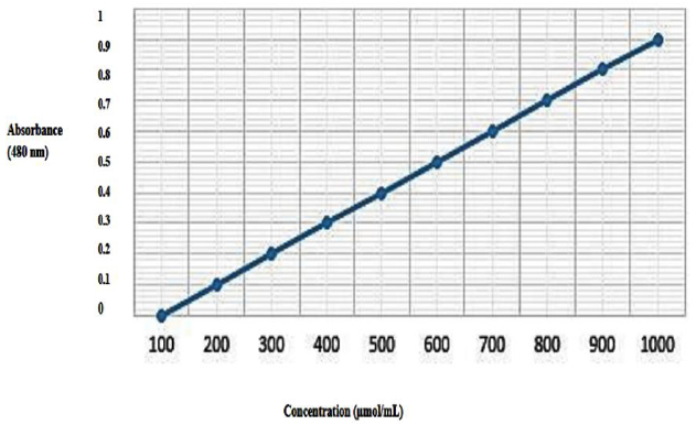
Standard ammonium sulfate absorbance curve.
3.3. Results of Partial Enzyme Purification
The purification of asparaginase was performed using ammonium sulfate precipitation and ion exchange chromatography. The specific activity of the enzyme after partial purification reached about 415.25 mg protein/unit, which indicates a significant increase in the purity of the enzyme (Table 3).
| Purification Stage | Specific Activity (mg protein/unit) | Fold Increase |
|---|---|---|
| Crude enzyme/before purification | 18.77 | 1.00 |
| Partially purified enzyme | 415.25 | 22.12 |
3.4. SDS-PAGE Results
Based on the SDS-PAGE electrophoresis pattern of the purified asparaginase, there was a dominant protein band with a molecular weight of ~30 kDa (Fig. 2). A comparison of specific activity before and after purification indicated that the relative purity of the enzyme increased about 21-fold during the ammonium sulfate precipitation step.
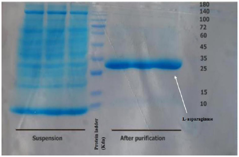
SDS-PAGE results show enzyme purification alongside a 180 kDa protein ladder.
3.5. Cell Proliferation Assay (MTT) Results
The MTT assay was used to assess the cytotoxic effect of the extracted asparaginase on the MCF-7 BC cell line, after 24 and 48 h of treatment (Fig. 3). The results exhibited both dose- and time-dependent reduction in cell viability after exposure to varying asparaginase concentrations. Unlike the untreated control group, the asparaginase-treated group significantly prevented MCF-7 cell proliferation. The IC50 values determined were 25.46 μg/mL after 24 h and 16.54 μg/mL after 48 h of treatment, which indicates increased cytotoxic activity of the enzyme with long-term exposure. These observations show that the extracted asparaginase has potential antiproliferative effects against MCF-7 BC cells.
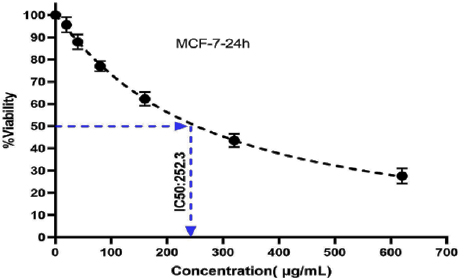
MTT assay result showing that the asparaginase extracted in this research significantly destroys cancer cells.
3.6. Results of the Complementary Identification of L-asparaginase-producing Strains
PCR results showed that a fragment of approximately 1500 bp from the 16S rRNA gene and a fragment of approximately 216 bp from the L-asparaginase gene were amplified, which were observed on 1% and 2% agarose gels, respectively (Fig. 4). The PCR products were purified and sent for sequencing. The obtained sequences were examined using CLC Sequence Viewer and FinchTV software and compared with the sequences available in the NCBI database via BLAST. The results showed high similarity to reference sequences. The phylogenetic position of the strain was determined after drawing a phylogenetic tree based on the 16S rRNA gene sequence and using the Neighbor-Joining method in MEGA X software. The studied strain was placed in the actinomycete group and had 100% similarity to Streptomyces flavoviridis (KP260508.1), which confirmed the final identification of the strain as Streptomyces flavoviridis (Fig. 5). The reference sequences used for the phylogenetic analysis are detailed in Table 4.
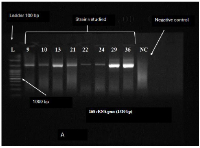
Electrophoresis of PCR product. (A) 16S rRNA gene (1520 bp); (B) the asparaginase gene (216 bp).
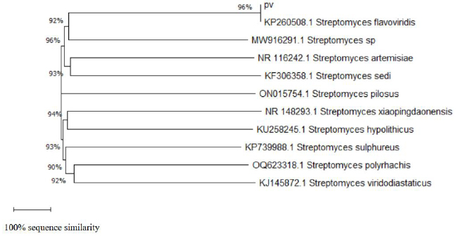
Phylogenetic dendrogram.
| Strain no. | Reference Species | Genbank Accession Number |
|---|---|---|
| 1 | Streptomyces flavoviridis | KP260508.1 |
| 2 | Streptomyces sp. | MW916291.1 |
| 3 | Streptomyces artemisiae | NR_116242.1 |
| 4 | Streptomyces sedi | KF306358.1 |
| 5 | Streptomyces pilosus | ON015754.1 |
| 6 | Streptomyces xiaopingdaonensis | NR_148293.1 |
| 7 | Streptomyces hypolithicus | KU258245.1 |
| 8 | Streptomyces sulphureus | KP739988.1 |
| 9 | Streptomyces polyrhachis | OQ623318.1 |
| 10 | Streptomyces viridodiastaticus | KJ145872.1 |
4. DISCUSSION
The aim of this research was to isolate the actinomycetes producing L-asparaginase from soil samples gathered from Qom City, optimize the production of L-asparaginase, and determine its anticancer effects in vitro. Actinomycetes, soil-dwelling bacteria, can produce secondary metabolites and industrial enzymes. L-asparaginase is one of the major anticancer drugs that has been applied for the treatment of acute lymphoblastic leukemia, but optimization of its production process is a biotechnological challenge [7, 20].
For the present study, the samples were collected from soil from different locations of Qom City, and the actinomycetes strains were isolated. Their ability to produce enzymes, optimal production conditions, and anticancer effects were assessed in the study. Using PCR and 16S rRNA gene sequencing, the selected strains were identified. According to the phylogenetic analysis, the initially isolated strain was found to be 100% similar to Streptomyces flavoviridis. The innovative aspects of our research entail the use of a native source, soil from Qom, to isolate L-asparaginase-producing actinomycetes, the combination of molecular and biochemical methods for identification of strains and evaluation of enzyme activity, and the optimization of enzyme production conditions at the laboratory scale. This optimization focused on environmental and nutritional parameters to enhance efficiency. Our study also investigated the in vitro anticancer effects of the enzyme L-asparaginase, allowing for an assessment of the therapeutic potential of native strains.
A previous study by Yousefi et al. (2019) primarily focused on the isolation and characterization of soil actinomycetes and their antibacterial activity or initial optimization of L-asparaginase production [21]. In contrast, our study combined molecular identification, production optimization, and a thorough investigation of anticancer effects, presenting a comprehensive and innovative approach. It demonstrates that native Iranian soil can serve as a rich and efficient source for the production of industrial and therapeutic enzymes. The study by Baraka et al. (2023) aimed to isolate and optimize asparaginase from actinomycetes [22]. They isolated 34 actinomycetes from soil and water in different regions of Egypt, identifying nine Streptomyces strains with high potential for L-asparaginase production. Their results showed that glucose and peptone were suitable carbon and nitrogen sources for optimal production efficiency, with optimal enzyme activity occurring at pH 7, a temperature of about 30 °C, and a seven-day incubation period. These findings indicate that optimizing environmental parameters can significantly increase enzyme activity and production, consistent with results from our study. Gopalakrishnan et al. (2024) evaluated the optimization of asparaginase from forest actinomycetes in India [23]. They observed that not only physical but also chemical parameters (pH ~8, temperature ~30 °C, and using glucose peptone as a food source) increased the production of asparaginase. The isolated enzyme demonstrated anticancer and antioxidant effects, underscoring its multidimensional potential for clinical and industrial applications. Their approach to isolating actinomycete strains from natural environments and optimizing physical/chemical parameters stated above led to significant production of L-asparaginase. Ali and associates (2025) demonstrated the recombinant production of L-asparaginase from Paenibacillus thiaminolyticus, achieving a yield of ~335 mg/L. This enzyme notably lacked L-glutaminase activity, which could reduce side effects in therapeutic applications. The enzyme exhibited stable biochemical properties, indicating its suitability for use in biosensors designed to measure L-asparagine in both therapeutic and industrial settings. The same authors optimized the extracellular production of L-asparaginase from Aspergillus oryzae through chemical mutagenesis. They initially screened fungal strains isolated from soil to assess their capacity for enzyme production, ultimately selecting a superior strain. Next, they optimized various culture conditions, including substrate type, moisture content, inoculum size, and incubation time, within a solid-state fermentation system. Notably, they used nitrous acid as a mutagenic agent to create mutant strains with enhanced enzyme activity. The results showed that the L-asparaginase activity in the mutant strain increased more than twofold compared to the parent strain, with the addition of certain mineral salts further boosting enzyme activity [24].
Overall, our study shows that combining the optimization of culture conditions with chemical mutagenesis is an effective method for increasing L-asparaginase production in fungi. This method can be used as a practical strategy for improving the efficiency of industrial production of this enzyme. The findings from this study and previous studies show improvements in biological and biochemical conditions for production and many other applications of asparaginase, which can lead to the development of therapeutic and industrial applications. In addition, this study confirms the high potential of indigenous fungal strains for pharmaceutical and industrial applications, paving the way for future research in industrial production of anticancer enzymes sourced from local microorganisms. However, it is important to note the limitations of this study. The anticancer effects of the enzyme were evaluated on a specific cell line, and the research was limited to a particular geographical area. Likewise, we did not conduct further studies to explore the molecular mechanisms underlying the observed cytotoxic effects.
CONCLUSION
Soil actinomycetes of Qom city have a potential capacity in the production of industrial enzymes, especially L-asparaginase. In addition to its medicinal use as an anticancer agent, this enzyme can also play an effective role in reducing carcinogens in the food industry. Optimizing fermentation conditions can lower production costs and improve enzyme yield, facilitating further research into reducing immunogenic side effects in subsequent studies.
AUTHORS’ CONTRIBUTIONS
The authors confirm their contributions to the paper as follows: study conception and design: M.R.Z. data collection: A.P.; analysis and interpretation of results: A.M.; draft manuscript: P.SH. All authors have reviewed the results and approved the final version of the manuscript.
LIST OF ABBREVIATIONS
| SDS-PAGE | = Sodium Dodecyl Sulfate-Polyacrylamide Gel Electrophoresis |
| BC | = Breast cancer |
| MTT | = Diphenyltetrazolium bromide |
AVAILABILITY OF DATA AND MATERIALS
The data and supporting information is available within the article.
ACKNOWLEDGEMENTS
The authors would like to thank the Vice Chancellor for Research, Islamic Azad University, Qom Branch, Qom, Iran.